中国组织工程研究 ›› 2020, Vol. 24 ›› Issue (32): 5168-5172.doi: 10.3969/j.issn.2095-4344.2866

• 组织构建实验造模 experimental modeling in tissue construction • 上一篇    下一篇

免疫性卵巢早衰模型鼠制备的时间

田海清,张于念,张和江,范莹露,腊晓琳   

  1. 新疆医科大学第一附属医院生殖医学中心,新疆维吾尔自治区乌鲁木齐市  830000
  • 收稿日期:2019-10-24 修回日期:2019-10-30 接受日期:2019-12-19 出版日期:2020-11-18 发布日期:2020-09-25
  • 通讯作者: 腊晓琳,博士,教授,新疆医科大学第一附属医院生殖医学中心,新疆维吾尔自治区乌鲁木齐市 830000
  • 作者简介:田海清,女,1976年生,山东省昌乐县人,汉族,2011年新疆医科大学毕业,硕士,副主任医师,主要从事生殖医学研究。
  • 基金资助:
    新疆维吾尔自治区自然科学基金(2017D01C318)

Time for preparing a mouse model of autoimmune premature ovarian failure

Tian Haiqing, Zhang Yunian, Zhang Hejiang, Fan Yinglu, La Xiaolin   

  1. Center for Reproductive Medicine, First Affiliated Hospital of Xinjiang Medical University, Urumqi 830000, Xinjiang Uygur Autonomous Region, China
  • Received:2019-10-24 Revised:2019-10-30 Accepted:2019-12-19 Online:2020-11-18 Published:2020-09-25
  • Contact: La Xiaolin, MD, Professor, Center for Reproductive Medicine, First Affiliated Hospital of Xinjiang Medical University, Urumqi 830000, Xinjiang Uygur Autonomous Region, China
  • About author:Tian Haiqing, Master, Associate chief physician, Center for Reproductive Medicine, First Affiliated Hospital of Xinjiang Medical University, Urumqi 830000, Xinjiang Uygur Autonomous Region, China
  • Supported by:
    the Natural Science Foundation of Xinjiang Uygur Autonomous Region, No. 2017D01C318

摘要:

文题释义:

免疫性卵巢早衰近年来卵巢早衰在育龄女性中的发病率逐年上升,且向低龄化发展, 成为女性不孕的常见重要原因之一。卵巢早衰的病因复杂,很多患者(50%-60%)找不到明确的原因,迄今为止卵巢早衰的发病机制不明,临床研究显示10%-30%的卵巢早衰是由免疫因素所致,其早期诊断困难,治疗也相当棘手。因此对于免疫导致卵巢早衰的研究,已成为目前国内外研究的热点。

免疫性卵巢早衰模型:免疫性卵巢早衰动物模型的建立为临床研究奠定了基础,因此需要一个建模方法简单,成功率较高,而且卵巢的形态和功能与人类卵巢早衰相似的动物模型。

背景:研究已经证实抗透明带抗体可以加速卵母细胞的破坏和耗竭而致卵巢早衰。
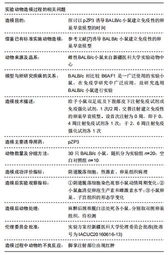
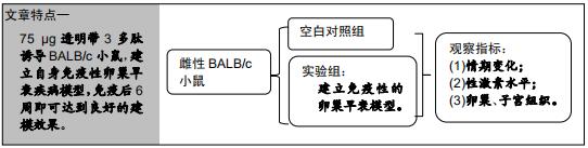
目的探讨以透明带3多肽(pZP3)诱导BALB/c小鼠建立免疫性的卵巢早衰模型的时间。
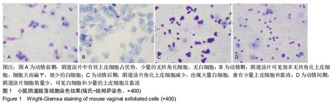
方法6-8周龄的健康雌性BALB/c小鼠30只,随机分为免疫实验组(20)和空白对照组(10)。实验组小鼠首次免疫注射时间为0周,给予0.15 mL免疫试剂注射双足底及下腹部皮下,每日晨阴道脱落细胞涂片观察小鼠动情周期的变化;2周后给予0.15 mL免疫强化试剂皮下注射相同部位,第4周和第6周的第1天,分别注射免疫试剂或免疫强化试剂各1(交替注射)。在每次注射前采血用酶联免疫法测小鼠血清性激素;最后观察小鼠卵巢组织及子宫形态。

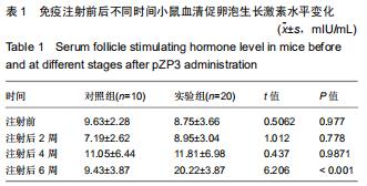
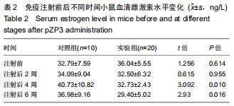
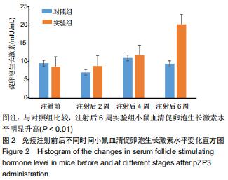
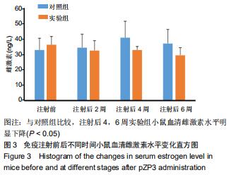
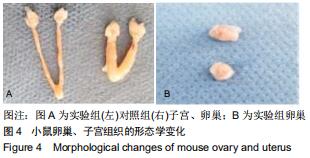
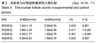
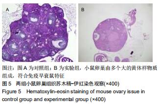
结果与结论:①实验组小鼠初次免疫注射及注射后2周,2组间血清促卵泡生长激素及雌激素水平差异无显著性意义;注射后4周实验组血清雌激素水平明显低于对照组,但促卵泡生长激素水平差异无显著性意义;注射后6周实验组血清雌激素水平明显低于对照组(P < 0.05),促卵泡生长激素水平明显高于对照组(P < 0.01);②实验组小鼠卵巢间质纤维化程度较对照组明显,卵巢体积减小,子宫萎缩,小鼠原始卵泡、初级及次级卵泡数均显著低于对照组,闭锁卵泡数量高于对照组(P < 0.05);③结论:75 μg透明带3多肽诱导BALB/c小鼠,建立自身免疫性卵巢早衰疾病模型,免疫后6周即可达到良好的建模效果。

ORCID: 0000-0001-8035-616X(田海清)

中国组织工程研究杂志出版内容重点:组织构建;骨细胞;软骨细胞;细胞培养;成纤维细胞;血管内皮细胞;骨质疏松;组织工程

关键词: 透明带3多肽, 鼠, 免疫性疾病, 卵巢早衰, 动物, 模型

Abstract:

BACKGROUND: Studies have confirmed that anti-zona zona antibodies can accelerate the destruction and depletion of oocytes, thereby causing premature ovarian failure.

OBJECTIVE: To investigate the time of establishing autoimmune premature ovarian failure model in BALB/c mice induced by zona pellucida 3 peptides.

METHODS: Thirty healthy female BALB/c mice (7-8 weeks) were randomly divided into immune experimental group (20 mice) and the control group (10 mice). In the experimental group, the mice were given immunization injection starting at 0 week, 0.15 mL of immune reagent injected into the soles at both sides and the lower

abdomen. The vaginal exfoliated cell smears were observed every morning to observe the changes in the estrous cycle of the mice. After 2 weeks of injection, 0.15 mL of immune enhancement agent was injected subcutaneously into the same site. On the first day of weeks 4 and 6, the immune agent or immune enhancement agent was injected alternatively. Blood samples were collected before each injection and the serum sex hormone level was measured by enzyme-linked immunosorbent assay. Finally, ovarian tissue and uterus morphology were observed.

RESULTS AND CONCLUSION: There was no difference in serum follicle stimulating hormone and estrogen levels between the first immunization injection and 2 weeks after the injection. The serum estrogen level in the experimental group was significantly lower than that in the control group at 4 weeks after injection. The serum estrogen level of the experimental group was significantly lower than that of the control group at 6 weeks after injection (P < 0.05), and meanwhile the serum follicle stimulating hormone level was significantly higher than that of the control group (P < 0.01). The degree of ovarian interstitial fibrosis in the experimental group was more obvious than that in the control group. The ovarian volume decreased and the uterus atrophied in the experimental group. The number of primitive follicles, primary follicles and secondary follicles in mice was significantly lower in the experimental group than the control group, and the number of atresia follicles was significantly higher than the control group (P < 0.05). Therefore, 75 μg of zona pellucida 3 peptides can be used to establish an autoimmune premature ovarian failure disease model in BALB/c mice, and a good modelling effect can be achieved at 6 weeks after immunization.

Key words: zona pellucida 3 peptides, mouse, immune disease, premature ovarian failure, animal, model

中图分类号: