中国组织工程研究 ›› 2020, Vol. 24 ›› Issue (20): 3130-3134.doi: 10.3969/j.issn.2095-4344.2611

• 骨组织构建 bone tissue construction • 上一篇    下一篇

脉冲电磁场磁疗与骨硬化蛋白单克隆抗体联合干预绝经后新西兰大白兔骨代谢和骨微结构的变化

钱  光,余月明,董有海,洪  洋,王明海   

  1. 复旦大学附属上海市第五人民医院骨科,上海市  200240
  • 收稿日期:2019-08-31 修回日期:2019-09-02 接受日期:2019-10-15 出版日期:2020-07-18 发布日期:2020-04-10
  • 通讯作者: 王明海,博士,主任医师,副教授,复旦大学附属上海市第五人民医院骨科,上海市 200240
  • 作者简介:钱光,男,1979年生,河南省睢县人,回族,2005年同济大学毕业,硕士,副主任医师,主要从事脊柱外科和组织工程研究。
  • 基金资助:
    上海市闵行区自然科学基金项目资助(2018MHZ038)

Pulsed electromagnetic fields stimulation combined with sclerostin antibody improves bone metabolism and bone microstructure in postmenopausal osteoporosis rabbits

Qian Guang, Yu Yueming, Dong Youhai, Hong Yang, Wang Minghai   

  1. Department of Orthopedics, Shanghai Fifth People’s Hospital, Fudan University, Shanghai 200240, China
  • Received:2019-08-31 Revised:2019-09-02 Accepted:2019-10-15 Online:2020-07-18 Published:2020-04-10
  • Contact: Wang Minghai, MD, Chief physician, Associate professor, Department of Orthopedics, Shanghai Fifth People’s Hospital, Fudan University, Shanghai 200240, China
  • About author:Qian Guang, Master, Associate chief physician, Department of Orthopedics, Shanghai Fifth People’s Hospital, Fudan University, Shanghai 200240, China
  • Supported by:
    the Natural Science Foundation of Minhang District of Shanghai, No. 2018MHZ038

摘要:

文题释义:

低频脉冲电磁场(pulsed electromagnetic fields,PEMFs)疗法:是目前常用的骨质疏松症物理治疗方法。脉冲电磁场治疗骨质疏松症的原理是采用低频脉冲电磁场改变人体生物电、改善生物场,促使成骨细胞增生,增强成骨能力,提高骨密度治疗骨质疏松。

骨硬化蛋白的单克隆抗体(sclerostin antibody,Scl-Ab):对骨代谢的影响集中体现在对骨硬化蛋白的拮抗作用上。骨硬化蛋白单克隆抗体在刺激成骨活动的同时,不会刺激破骨活动,对骨合成代谢有显著刺激作用,其已经成为治疗骨质疏松的潜在方法。

背景:脉冲电磁场与骨硬化蛋白单克隆抗体皆能对绝经后新西兰大白兔骨代谢产生良好影响,但关于两者联合干预的效果至今少有报道。

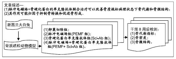
目的:探讨脉冲电磁场联合骨硬化蛋白单克隆抗体对绝经后新西兰大白兔骨代谢的影响,探索其对骨质疏松症的治疗价值。

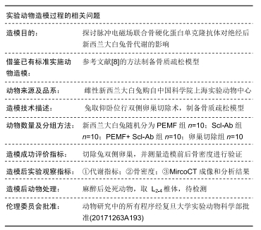
方法:采用卵巢切除法建立新西兰大白兔绝经后骨质疏松动物模型。将实验动物随机分为4组:卵巢切除组、脉冲电磁场组、骨硬化蛋白单克隆抗体组和脉冲电磁场+骨硬化蛋白单克隆抗体组,每组10只。术后第1天起,脉冲电磁场组给予脉冲电磁场磁疗每天1次;骨硬化蛋白单克隆抗体组给予骨硬化蛋白单克隆抗体皮下注射每周2次;脉冲电磁场+骨硬化蛋白单克隆抗体组接受脉冲电磁场磁疗每天1次、每周5次,骨硬化蛋白单克隆抗体皮下注射每周2次;卵巢切除组皮下注射相同剂量的生理盐水每周2次,干预共8周。8周后行骨代谢指标检查、骨密度测定、MicroCT骨微结构参数检测。动物研究中的所有程序经复旦大学实验动物科学部批准(20171263A193)。

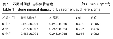
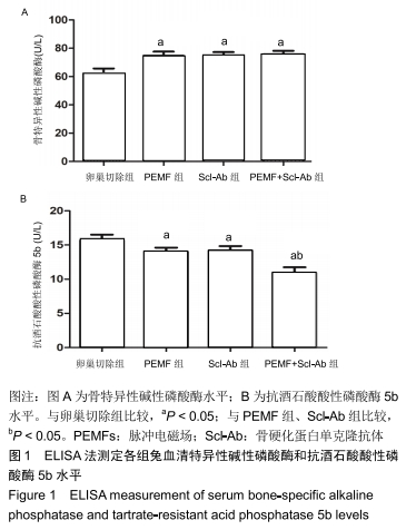
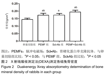
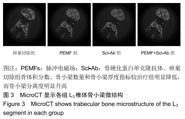
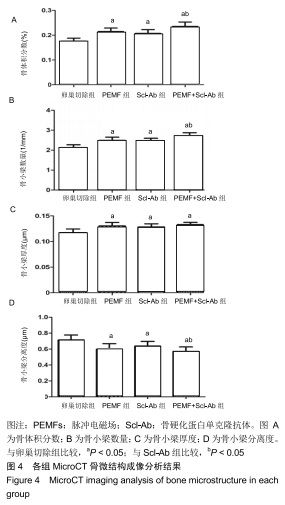
结果与结论:①卵巢切除6个月新西兰大白兔骨密度显著下降,提示骨质疏松模型建立成功;②与卵巢切除组相比,3个治疗组L3椎体的骨密度均显著增加(P < 0.05);③3个治疗组血清骨特异性碱性磷酸酶水平均显著高于卵巢切除组,血清抗酒石酸酸性磷酸酶5b水平均显著低于卵巢切除组;脉冲电磁场+骨硬化蛋白单克隆抗体组血清抗酒石酸酸性磷酸酶5b的水平明显低于脉冲电磁场组、骨硬化蛋白单克隆抗体组;④脉冲电磁场+骨硬化蛋白单克隆抗体组骨微结构参数(骨体积分数、骨小梁厚度、骨小梁数量、骨小梁分离度)均优于脉冲电磁场组、骨硬化蛋白单克隆抗体组(均P < 0.05);⑤骨硬化蛋白单克隆抗体和脉冲电磁场联合治疗可以增强去势新西兰大白兔骨密度,改善骨代谢和骨微结构。

ORCID: 0000-0001-7052-4262(钱光)

中国组织工程研究杂志出版内容重点:组织构建;骨细胞;软骨细胞;细胞培养;成纤维细胞;血管内皮细胞;骨质疏松组织工程

关键词:

脉冲电磁场, 骨硬化蛋白单克隆抗体, 联合治疗, 骨质疏松症, 骨量, 骨密度,

Abstract:

BACKGROUND: Both pulsed electromagnetic fields (PEMF) stimulation and sclerostin antibody (Scl-Ab) have good effects on the bone metabolism of ovariectomized (OVX) New Zealand rabbits, but research on the combined intervention of PEMF and Scl-Ab in the OVX rabbits is rarely reported.

OBJECTIVE: To explore the effect of PEMF combined with Scl-Ab on postmenopausal osteoporosis and to explore the therapeutic value for osteoporosis.

METHODS: An animal model of postmenopausal osteoporosis was made in New Zealand white rabbits after ovariectomy. The experimental animals were randomly divided into OVX control group, PEMF group, Scl-Ab group and PEMF+Scl-Ab group, with 10 rats in each group. On the 1st day after surgery, the PEMF group was given PEMF magnetic therapy once a day; the Scl-Ab group was given subcutaneous injection of Scl-Ab twice a week; the PEMF+Scl-Ab group received PEMF magnetic therapy once a day, five times a week, and Scl-Ab subcutaneous injections twice a week; the OVX group was injected subcutaneously with the same dose of normal saline twice a week for 8 weeks. After 8 weeks of treatment, bone metabolism index, bone mineral density, and MicroCT bone microstructure parameters were detected. All animal procedures were approved by the Department of Experimental Animal Science, Fudan University (approval No. 20171263A193).

RESULTS AND CONCLUSION: Bone mineral density was significantly decreased in the New Zealand white rabbits after 6 months of OVX, suggesting that the osteoporosis model was successfully established. Compared with the OVX group, the bone mineral density of the L3 vertebral body in the PEMF group, the Scl-Ab group and the PEMF+Scl-Ab group increased significantly (P < 0.05). Compared with the OVX group, serum bone-specific alkaline phosphatase levels were significantly higher, and serum tartrate-resistant acid phosphatase 5b levels were significantly lower in the PEMF group, the Scl-Ab group and the PEMF+Scl-Ab group. The serum tartrate-resistant acid phosphatase 5b level in the PEMF+Scl-Ab group was significantly lower than that in the PEMF group and the Scl-Ab group. The bone metabolism index and bone microstructural parameters (bone volume fraction, trabecular thickness, trabecular number, and trabecular separation) of the PEMF+Scl-Ab group were significantly better than those of the PEMF group and the Scl-Ab group (all P < 0.05). These findings indicate that the combination of Scl-Ab and PEMF can enhance bone mineral density and improve bone metabolism and bone microstructure in postmenopausal osteoporosis New Zealand white rabbits.

Key words: pulsed electromagnetic field, sclerostin antibody, combination treatment, osteoporosis, bone mass, bone mineral density, rabbit

中图分类号: