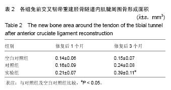
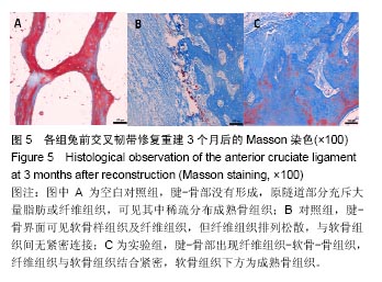
| [1]Clancy WG.Knee ligamentous injury in sports: the past, present, and future.Med Sci Sports Exerc.1983;15:9-14.[2]Filbay SR,Culvenor AG,Ackerman IN,et al.Quality of life in anterior cruciate ligament-deficient individuals: a systematic review and meta-analysis.Br J Sports Med. 2015;49: 1033-1041.[3]柴双,吕松岑.前交叉韧带重建术后促进腱-骨愈合因素的研究进展[J].医学综述,2017,23(4):718-723.[4]Chen B,Li B,Qi YJ,et al.Enhancement of tendon-to-bone healing after anterior cruciate ligament reconstruction using bone marrow-derived mesenchymal stem cells genetically modified with bFGF/BMP2.Sci Rep.2016;6:25940.[5]柴昉,蒋佳,陈世益.促进前交叉韧带腱骨愈合的生物治疗技术研究进展[J].中国运动医学杂志,2016,35(4):372-377.[6]张义龙,李宁,宋有鑫,等.自体浓缩骨髓移植促进腱骨愈合[J].中国组织工程研究,2013,17(2):223-227.[7]Nau T,Teuschl A.Regeneration of the anterior cruciate ligament: Current strategies in tissue engineering.World J Orthop.2015;6(1):127-136.[8]杨大志,罗惠莉,易伟宏,等.转染骨形态发生蛋白-2基因的人脐带间充质干细胞成骨研究[J].中华实验外科杂志, 2012,29(8): 1580-1583.[9]Yang J,Guan K,Wang JZ.Clinical study on the arthroscopic refreshing treatment of anterior cruciate ligament injury combined with stable medial meniscus ramp injury. JMusculoskelet Neuronal Interact.2017;17(2):108-113.[10]Beaulieu ML,Carey GE,Schlecht SH,et al.On the heterogeneity of the femoral enthesis of the human ACL: microscopic anatomy and clinical implications.J Exp Orthop. 2016;3(1):14.[11]Zhang L,Jiang K,Chai H,et al.A Comparative Animal Study of Tendon Grafts Healing After Remnant-Preserving Versus Conventional Anterior Cruciate Ligament Reconstruction.Med Sci Monit. 2016;22:3426-3437.[12]Thomopoulos S,Genin GM,Galatz LM.The development and morphogenesis of the tendon-to-bone insertion - What development can teach us about healing.J Musculoskelet Neuronal Interact.2010;10:35-45.[13]田明,余家阔,吴延平.反复冻融同种异体跟腱重建前交叉韧带移植物的组织学特点[J].中国组织工程研究, 2017,21(16): 2546-2551 .[14]Setiawati R,Utomo DN,Rantam FA,et al.Early Graft Tunnel Healing After AnteriorCruciate Ligament Reconstruction With Intratunnel Injection of Bone Marrow Mesenchymal Stem Cells and Vascular Endothelial Growth Factor.Orthop J Sports Med.2017;5(6):2325967117708548.[15]Chen CH.Graft healing in anterior cruciate ligament reconstruction.Sports Med Arthrosc Rehab Ther Tech. 2009;1:21.[16]Rodeo SA,Voigt C,Ma R,et al.Use of a new model allowing controlled uniaxial loading to evaluate tendon healing in a bone tunnel.J Orthop Res.2016;34(5):852-859.[17]Thomopoulos S,Zampiakis E,Das R,et al.The effect of muscle loading on flexor tendon-to-bone healing in a canine model.J Orthop Res.2008;26:1611-1617.[18]Howells BE,Clark RA,Ardern CL,et al.The assessment of postural control and the influence of a secondary task in people with anterior cruciate ligament reconstructed knees using a Nintendo Wii Balance Board.Br J Sports Med.2013; 47(14):914-919.[19]Cheng P,Han P,Zhao C,et al.High-purity magnesium interference screws promote fibrocartilaginousentheses regeneration in the anterior cruciate ligament reconstruction rabbit model via accumulation of BMP-2 and VEGF. Biomaterials. 2016;81:14-26.[20]纪庆明,杨育晖,陈昊,等.富血小板血浆辅助前交叉韧带重建治疗的临床疗效分析[J].中国修复重建外科杂志, 2017,31(4): 410-416.[21]Ardern CL,Taylor NF,Feller JA,et al.Psychological responses matter in returning to preinjury level of sport after anterior cruciate ligament reconstruction surgery.Am J Sports Med. 2013;41(7):1549-1558.[22]Hu J,Qu J,Xu D,et al.Combined application of low-intensity pulsed ultrasound and functional electrical stimulation accelerates bone-tendon junction healing in a rabbit model.J Orthop Res.2014;32(2):204-209[23]Isaac C,Gharaibeh B,Witt M,et al.Biologic approaches to enhance rotator cuff healing after injury. J Shoulder Elbow Surg.2012;21:181-190.[24]董向辉,凌鸣,冯伟楼,等.关节滑液对兔前交叉韧带重建后腱骨愈合生物力学和组织学的影响[J].中国组织工程研究, 2012,16(11): 1937-1940.[25]Teng C,Zhou C,Xu D,et al.Combination of platelet-rich plasma and bone marrow mesenchymal stem cells enhances tendon-bone healing in a rabbit model of anterior cruciate ligament reconstruction.J Orthop Surg Res.2016;11(1):96.[26]HaoZC,Wang SZ, Zhang XJ,et al.Stem cell therapy: a promising biological strategy for tendon-bone healing after anterior cruciate ligament reconstruction.Cell Prolif.2016; 49(2):154-162.[27]李爱国,闫军伟,陈鸿辉,等.Ⅰ型胶原-血管内皮生长因子缓释材料促进兔骨-肌腱结合部位愈合的作用[J].中华创伤杂志, 2015, 31(6):557-561.[28]Bi F,Shi Z,Liu A,et al.AnteriorCruciate Ligament Reconstruction in a Rabbit Model Using Silk-Collagen Scaffold and Comparison with Autograft.PLoS One. 2015; 10(5): e0125900.[29]Han F,Zhang P,Sun Y,et al.Hydroxyapatite-doped polycaprolactone nanofiber membrane improves tendon–bone interface healing for anteriorcruciate ligament reconstruction.Int J Nanomedicine.2015;10:7333-7343. [30]Manning CN,Kim HM,Sakiyama-Elbert S,et al.Sustained delivery of transforming growth factor beta three enhances tendon-to bone healing in a rat model.J Orthop Res. 2011; 29(7):1099-1105.[31]Dai Z,Li S,Bao W,et al.Enhancement of polyethylene terephthalate artificial ligament graft osseointegration using a periosteum patch in a goat model.Int J Sports Med. 2016; 37(6):493-499.[32]Chou YC,Yeh WL,Chao CL,et al.Enhancement of tendon–bone healing via the combination of biodegradable collagen-loaded nanofibrous membranes and a three-dimensional printed bone-anchoring bolt.Int J Nanomedicine.2016;11:4173-4186.[33]杨大志,贺永进,郭晓东,等.骨形态发生蛋白-2对人脐带间充质干细胞增殖和分化的影响[J].中华实验外科杂志, 2010,27(11): 1602-1605.[34]Martinek V,Latterman C,Usas A,et al.Enhancement of tendon-bone integration of anterior cruciate ligament grafts with bone morphogenetic protein-2 gene transfer: a histological and biomechanical study.J Bone Joint Surg Am.2002;84-A(7):1123-1131.[35]Dong Y,Zhang Q,Li Y,et al. Enhancement of tendon-bone healing for anterior cruciate ligament (ACL) reconstruction using bone marrow-derived mesenchymal stem cells infected with BMP-2.Int J Mol Sci. 2012;13(10):13605-13620.[36]Lee KW,Lee JS,Jiang JW,et al.Tendon–bone interface healing using an injectable rhBMP-2-containing collagen gel in a rabbit extra-articular bone tunnel model.J Tissue Regen Med. 2017;11:1435-1441.[37]Takigami J,Hashimoto Y,Yamasaki S,et al.Direct bone-to-bone integration between recombinant human bone morphogenetic protein-2-injected tendon graft and tunnel wall in an anterior cruciate ligament reconstruction model.Int Orthop.2015;39(7):1441-1447. [38]Chen B,Li B,Qi YJ,et al.Enhancement of tendon-to-bone healing after anterior cruciate ligament reconstruction using bone marrow-derived mesenchymal stem cells genetically modified with bFGF/BMP2.Sci Rep.2016;6:25940.[39]Schwarting T,Schenk D,Frink M,et al.Stimulation with bone morphogenetic protein-2 (BMP-2) enhances bone–tendon integration in vitro.Connect Tissue Res.2016;57(2):99-112. |
.jpg)
.jpg)
.jpg)