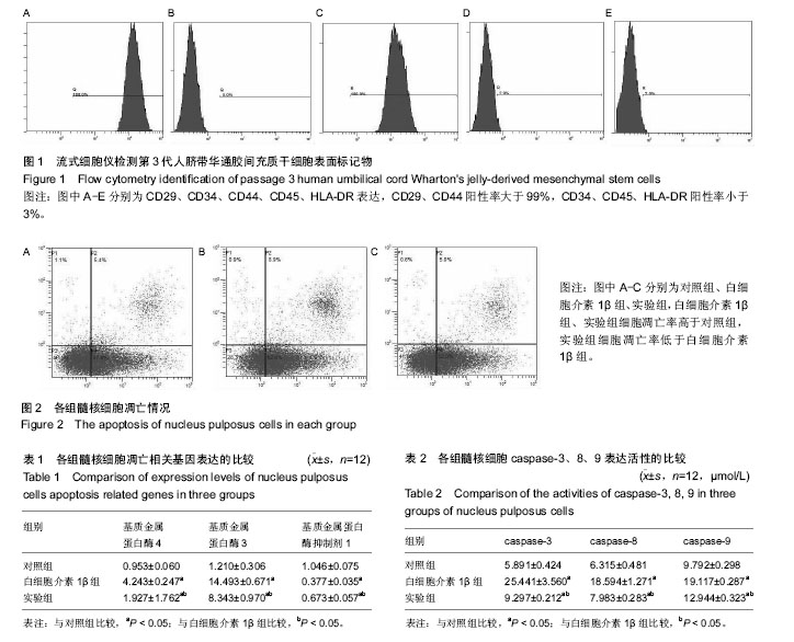
| [1] Buchbinder R,Blyth FM,March LM,et al.Placing the global burden of low back pain in context.Best Pract Res Clin Rheumatol.2013;27:575-589.[2] Vos T,Flaxman AD,Naghavi M,et al.Years lived with disability (YLDs)for 1160 sequelae of 289 diseases and injuries 1990-2010: a systematic analysis for the Global Burden of Disease Study 2010.Lancet.2012;380:2163-2196. [3] Freemont AJ.The cellular pathobiology of the degenerate intervertebral disc and discogenic back pain. Rheumatology (Oxford).2009;48(1):5-10.[4] Hoyland JA,Le Maitre C,Freemont AJ.nvestigation of the role of IL-1 and TNF in matrix degradat ion in the intervertebral disc.Rheumato logy(Oxford).2008;47(6):809-814. [5] Le Maitre CL,Freemont AJ,Hoyland JA.The role of interleukin-1 in the pathogenesis of human intervertebral disc degeneration.Arth Res Ther.2005;7(4):1-14.[6] Zhang Y,Tao H,Gu T,et al.The effects of human Wharton’s jelly cell transplantation on the intervertebral disc in a canine disc degeneration model.Stem Cell Res Ther.2015;6(1):154. [7] Zhang Y,Ruan D,Zhang C,et al.Differentiation of human umbilical cord Wharton’s jelly—derived mesenchymaI stem cells into nucleuspulposus-like cells by coculturetll cell-cell contact in vitro.Chin J Spine Spinal Cord.2012; 22(10): 936-942.[8] Gou S,Oxentenko SC,Eldrige JS,et al.Stem cell therapy for intervertebral disk regeneration.Am J Phys Med Rehabil. 2014;93:S122-131.[9] Mazzini L,Mareschi K,Ferrero I,et al.Mesenchyal stem cell transplantation in amyotrophic lateral sclerosis:a long-term safety study.Cytotherapy.2012;14(1):56-60. [10] Zheng X,Xia C,Chen Z,et al.Requirement of the phosphatidylinositol 3-kinase/Akt signaling pathway for the effect of nicotine on interleukin-1beta-induced chondrocyte apoptosis in a rat model of osteoarthritis.Biochem Biophys Res Commun.2012;423(3):606-612.[11] Hu JQ,Deng GY,Tian Y,et al.An in vitro investigation into the role of bone marrow-derived mesenchymal stem cells in the control of disc degeneration.Mol Med Rep.2015;12(4): 5701-5708.[12] Mueller SM,Glowacki J.Age-related decline in the osteogenic potential of human bone marrow cells cultured in three-dimensional collagen sponges.J Cell Biochem.2001; 82(4):583-590.[13] Lee SY,Miwa M,Sakai Y,et al.In vitro multipotentiality and characterization of human unfractured traumatic hemarthrosis-derived progenitor cells: A potential cell source for tissue repair.J Cell Physiol.2007;210(3):561-566.[14] Wang L,Tran I.A Comparison of Human Bone Marrow–Derived Mesenchymal Stem Cells and Human Umbilical Cord–Derived Mesenchymal Stromal Cells for Cartilage Tissue Engineering.Tissue Eng.2009;15(18): 2259-2266.[15] Sobolewski K,Bankowski E,Chyczewski L.Collagen and glycosaminoglycans of Wharton’jclly.Biol Neonale.1997; 71(1):11-21.[16] Kulyk WM,Franklin JL,Hoffman LM.Sox9 expression during chondrogenesis in micromass cultures of embryonic linb mesenchymal.Exp Cell Res.2000;255(2):327-332.[17] Li C,Chen X,Qiao S.Effects of Wharton's jelly cells of the human umbilical cord on acute spinal cord injury in rats, and expression of interleukin-1βand nerve growth factor in spinal cord tissues.Artif Cells Nanomed Biotechnol.2016; 44(5): 1254-1258.[18] Fang TC,Pang CY,Chiu SC,et al.Renoprotective effect of human umbilical cord-derived mesenchymal stem cells in immunodeficient mice suffering from acute kidney injury.PLoS One.2012;7(9):e46504.[19] Ruiz WC,Malandrino A,Van Rijsbergen MM.Simulating the sensitivity of cell nutritive environment to composition changes within the intervertebral disc.J Mech Phys Solids. 2016;90:108-123.[20] Korecki CL,MacLean JJ,Iatridis JC.Dynamic compression effects on intervertebral disc mechanics and biology. Spine(Phila Pa 1976).2008;33(13):1403-1409. [21] Saliken DJ,Mulet-Sierra A,Jomha NM,et al.Decreased hypertrophic differentiation accompanies enhanced matrix formation in co-cultures of outer meniscus cells with bone marrow mesenchymal stromal cells.Arthritis Res Ther.2012; 14(3):R153.[22] Gardner J,Ghorpade A.Tissue inhibitor of metalloproteinase (TIMP)-1: the TIMPed balance of matrix metalloproteinases in the central nervous system.J Neurosci Res.2003;74(6):801- 806.[23] Erwin WM,Islam D,Inman RD,et al.Notochordal cells protect nucleus pulposus cells from degradation and apoptosis: implications for the mechanisms of intervertebral disc degeneration.Arthritis Res Ther.2011;13(6):R215.[24] Liang S,Sun K,Wang Y,et al.Role of Cyt-C/caspases-9,3,Bax/Bcl-2 and the FAS death receptor pathway in apoptosis induced by zinc oxide nanoparticles in human aortic endothelial cells and the protective effect by alpha-lipoic acid.Chem Biol Interact.2016;25(258):40-51.[25] French LE,Tsehopp J.Protein-based therapeutic approaches targeting death receptors.Cell Death Differ.2003;10(1): 117-123.[26] Wajant H.Death receptors.Essays Biochem.2003;39:53-71.[27] Yamada K,Sudo H,Iwasaki K,et al.Caspase-3 silencing inhibits biomechanical overload-induced intervertebral disk degeneration.Am J Pathol.2014;184(3):753-764.[28] Division C.Department of Medicine. Paracrine mechanisms of Mesenchymal Stem cell-based therapy: Current status and perspectives.Cell Transplant.2014;23(9):1045-1059. |
.jpg)
.jpg)
.jpg)