中国组织工程研究 ›› 2016, Vol. 20 ›› Issue (51): 7622-7627.doi: 10.3969/j.issn.2095-4344.2016.51.003
• 骨组织构建 bone tissue construction • 上一篇 下一篇
MAPK抑制剂对骨碎补总黄酮促进转基因成肌细胞成骨分化的影响
张 力1,徐勃兴1,李 杰2,王 伟3
- 1锦州医科大学锦州临床学院/锦州市中心医院骨科,辽宁省锦州市 121000;锦州医科大学,2研究生学院,3骨外科学研究所,辽宁省锦州市 121000
Influence of mitogen-activated protein kinase inhibitor in the process of assemble flavone of rhizome drynaria promoting the osteogentic differentiation of myoblasts
Zhang Li1, Xu Bo-xing1, Li Jie2, Wang Wei3
- 1Department of Orthopaedics, Jinzhou Central Hospital of Jinzhou Medical University, Jinzhou 121000, Liaoning Province, China; 2Graduate School of Jinzhou Medical University, Jinzhou 121000, Liaoning Province, China; 3Institute for Orthopaedics, Jinzhou Medical University, Jinzhou 121000, Liaoning Province, China
摘要:
文章快速阅读:
.jpg) 文题释义:
水龙骨科:陆生或附生蕨类植物;根状茎横走,被阔鳞片;网状中柱;叶同型或二型;叶柄与根状茎有关节相连;单叶,全缘或羽状半裂至一回羽状分裂;网状脉。孢子囊群圆形或线形,或有时布满叶背,无囊群盖;孢子囊梨形或球状梨形;孢子两面形。
丝裂原活化蛋白激酶(mitogen-activated protein kinase,MAPK):是一组能被不同的细胞外刺激,如细胞因子、神经递质、激素、细胞应激及细胞黏附等激活的丝氨酸-苏氨酸蛋白激酶。由于MAPK是培养细胞在受到生长因子等丝裂原刺激时被激活而被鉴定的,因而得名。所有的真核细胞都能表达MAPK。MAPK通路的基本组成是一种从酵母到人类都保守的三级激酶模式,包括MAPK激酶激酶(MAP kinase kinase kinase,MKKK)、MAPK激酶(MAP kinase kinase,MKK)和MAPK,这3种激酶能依次激活,共同调节着细胞的生长、分化、对环境的应激适应、炎症反应等多种重要的细胞生理/病理过程。
文题释义:
水龙骨科:陆生或附生蕨类植物;根状茎横走,被阔鳞片;网状中柱;叶同型或二型;叶柄与根状茎有关节相连;单叶,全缘或羽状半裂至一回羽状分裂;网状脉。孢子囊群圆形或线形,或有时布满叶背,无囊群盖;孢子囊梨形或球状梨形;孢子两面形。
丝裂原活化蛋白激酶(mitogen-activated protein kinase,MAPK):是一组能被不同的细胞外刺激,如细胞因子、神经递质、激素、细胞应激及细胞黏附等激活的丝氨酸-苏氨酸蛋白激酶。由于MAPK是培养细胞在受到生长因子等丝裂原刺激时被激活而被鉴定的,因而得名。所有的真核细胞都能表达MAPK。MAPK通路的基本组成是一种从酵母到人类都保守的三级激酶模式,包括MAPK激酶激酶(MAP kinase kinase kinase,MKKK)、MAPK激酶(MAP kinase kinase,MKK)和MAPK,这3种激酶能依次激活,共同调节着细胞的生长、分化、对环境的应激适应、炎症反应等多种重要的细胞生理/病理过程。摘要
背景:前期研究中已证实低浓度骨碎补总黄酮含药血清对转睫状神经生长因子成肌细胞向成骨细胞分化具有促进作用,然而其潜在机制尚未明确。
目的:观察MAPK抑制剂对骨碎补总黄酮促进转基因成肌细胞成骨分化的影响。
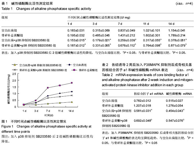
方法:转睫状神经生长因子成肌细胞预处理接种后,在成骨诱导剂诱导下,检测空白血清、骨碎补总黄酮含药血清及加入p38 MAPK抑制剂SB203580各组碱性磷酸酶比活性变化,RT-PCR检测成骨相关基因核结合因子a1和碱性磷酸酶mRNA表达的差异,并采用Western-blotting技术定量分析加入p38 MAPK抑制剂后成骨相关蛋白核结合因子a1和碱性磷酸酶蛋白表达情况的变化,进行统计学分析。
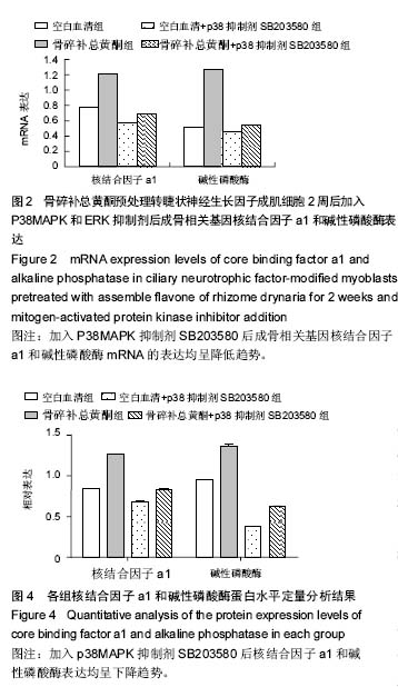
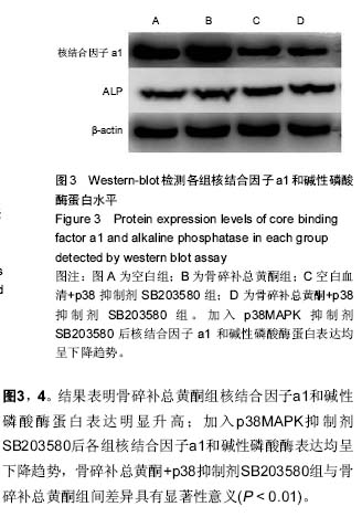
结果与结论:①RT-PCR检测结果均显示骨碎补总黄酮血清组的成骨相关因子核结合因子a1 mRNA、碱性磷酸酶mRNA表达最高,加入p38抑制剂SB203580后核结合因子a1 mRNA、碱性磷酸酶 mRNA表达明显下调;②Western-blot检测加入p38抑制剂SB203580后核结合因子a1 mRNA、碱性磷酸酶 mRNA蛋白水平明显下降;③结果说明,p38 MAPK抑制剂可以下调骨碎补总黄酮促进转基因成肌细胞成骨相关基因表达及蛋白水平,骨碎补总黄酮可能通过激活p38 MAPK信号通路促进睫状神经生长因子成肌细胞向成骨细胞分化。
中国组织工程研究杂志出版内容重点:组织构建;骨细胞;软骨细胞;细胞培养;成纤维细胞;血管内皮细胞;骨质疏松;组织工程
ORCID: 0000-0001-9259-4130(张力)
中图分类号:
.jpg) 文题释义:
水龙骨科:陆生或附生蕨类植物;根状茎横走,被阔鳞片;网状中柱;叶同型或二型;叶柄与根状茎有关节相连;单叶,全缘或羽状半裂至一回羽状分裂;网状脉。孢子囊群圆形或线形,或有时布满叶背,无囊群盖;孢子囊梨形或球状梨形;孢子两面形。
丝裂原活化蛋白激酶(mitogen-activated protein kinase,MAPK):是一组能被不同的细胞外刺激,如细胞因子、神经递质、激素、细胞应激及细胞黏附等激活的丝氨酸-苏氨酸蛋白激酶。由于MAPK是培养细胞在受到生长因子等丝裂原刺激时被激活而被鉴定的,因而得名。所有的真核细胞都能表达MAPK。MAPK通路的基本组成是一种从酵母到人类都保守的三级激酶模式,包括MAPK激酶激酶(MAP kinase kinase kinase,MKKK)、MAPK激酶(MAP kinase kinase,MKK)和MAPK,这3种激酶能依次激活,共同调节着细胞的生长、分化、对环境的应激适应、炎症反应等多种重要的细胞生理/病理过程。
文题释义:
水龙骨科:陆生或附生蕨类植物;根状茎横走,被阔鳞片;网状中柱;叶同型或二型;叶柄与根状茎有关节相连;单叶,全缘或羽状半裂至一回羽状分裂;网状脉。孢子囊群圆形或线形,或有时布满叶背,无囊群盖;孢子囊梨形或球状梨形;孢子两面形。
丝裂原活化蛋白激酶(mitogen-activated protein kinase,MAPK):是一组能被不同的细胞外刺激,如细胞因子、神经递质、激素、细胞应激及细胞黏附等激活的丝氨酸-苏氨酸蛋白激酶。由于MAPK是培养细胞在受到生长因子等丝裂原刺激时被激活而被鉴定的,因而得名。所有的真核细胞都能表达MAPK。MAPK通路的基本组成是一种从酵母到人类都保守的三级激酶模式,包括MAPK激酶激酶(MAP kinase kinase kinase,MKKK)、MAPK激酶(MAP kinase kinase,MKK)和MAPK,这3种激酶能依次激活,共同调节着细胞的生长、分化、对环境的应激适应、炎症反应等多种重要的细胞生理/病理过程。