中国组织工程研究 ›› 2016, Vol. 20 ›› Issue (16): 2391-2396.doi: 10.3969/j.issn.2095-4344.2016.16.015

• 药物控释材料 drug delivery materials • 上一篇    下一篇

高吸型壳聚糖敷料的创面止血及促愈合效果

毛 珺1,周应山2,吴 庭3   

  1. 1武汉纺织大学医院全科,湖北省武汉市 430073;2新型纺织材料绿色加工及其功能化省部共建教育部重点实验室,武汉纺织大学,湖北省武汉市 430073;3武汉市第三医院,湖北省武汉市 430073
  • 收稿日期:2016-02-11 出版日期:2016-04-15 发布日期:2016-04-15
  • 通讯作者: 吴庭,副主任医师,武汉市第三医院皮肤科,湖北省武汉市430073
  • 作者简介:毛珺,女,1981年生,湖北省武汉市人,汉族,硕士,主治医师,主要从事全科及相关临床研究。
  • 基金资助:

    2015年度湖北省教育厅重点项目(D20151605)

High-absorbing chitosan dressings for hemostasis and wound healing

Mao Jun1, Zhou Ying-shan2, Wu Ting3   

  1. 1Hospital of Wuhan Textile University, Wuhan 430073, Hubei Province, China; 2Key Laboratory for Green Processing and Functionalization of Textile Materials, Ministry of Education, Wuhan Textile University, Wuhan 430073, Hubei Province, China; 3Wuhan Third Hospital, Wuhan 430073, Hubei Province, China
  • Received:2016-02-11 Online:2016-04-15 Published:2016-04-15
  • Contact: Wu Ting, Associate chief physician, Wuhan Third Hospital, Wuhan 430073, Hubei Province, China
  • About author:Mao Jun, Master, Attending physician, Hospital of Wuhan Textile University, Wuhan 430073, Hubei Province, China
  • Supported by:

    the Key Project of Hubei Provincial Education Department in 2015, No. D20151605

摘要:

文章快速阅读:


文题释义:
高吸型壳聚糖敷料:是一种羧乙基甲壳胺纤维,将甲壳胺纤维分散在醇类中,通过与丙烯酸的迈克尔加成反应,经分离、洗涤、干燥得到羧乙基甲壳胺纤维。这种羧乙基甲壳胺纤维,羧化取代度控制在0.01-0.8范围内,羧化壳聚糖纤维分子链间的氢键被部分破坏,亲水基团大量引入,使得敷料中羧化壳聚糖纤维内部和纤维之间能吸收大量的渗出液,相对于一般甲壳胺纤维制备的常规壳聚糖敷料而言,提升其止血和吸液能力。
高吸型壳聚糖敷料修复烧伤创面:在炎症阶段,壳聚糖能激活巨噬细胞,促进其迁移,以达到清除伤口分泌物的作用;在肉芽阶段,壳聚糖能加速Ⅲ型胶原蛋白的分泌,从而促进肉芽组织的生长;在上皮化阶段,敷料中壳聚糖分子链在创面溶菌酶的作用下,降解产生的N-乙酰葡胺糖和葡糖胺烯糖小分子能被创面吸收,促进上皮细胞的再生,显著提高创面愈合速度和质量。高吸型壳聚糖分子链上定位接枝的羧化基团会给出质子,致使壳聚糖分子链上氨基质子化,增加壳聚糖纤维的聚阳离子特性,赋予其高抗菌性,相对以往壳聚糖敷料来说,促愈合、抗菌性有所提高。
 
背景:通过化学改性可提升壳聚糖敷料自身的高吸液性、高容量吸液性、高抗菌性等方面的性能。
目的:观察高吸型壳聚糖敷料的创面止血及促愈合效果。
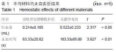
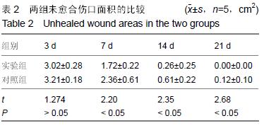
方法:①小静脉止血实验:制作家兔皮肤对称性渗血伤口,两侧伤口分别敷以高吸型壳聚糖敷料和灭菌纱布,记录止血时间和出血量;②Ⅱ度烫伤创面修复实验:取清洁级SD大鼠40只,制作背部深Ⅱ度烫伤创面,随机分为2组,实验组创面局部以高吸型壳聚糖敷料包扎,对照组以凡士林纱布包扎,修复后3,7,14,21 d,观察愈合情况,取创面组织进行病理组织学分析。
结果与结论:①小静脉止血实验:高吸型壳聚糖敷料组出血量与止血时间均低于灭菌纱布组(P < 0.05,P < 0.01);②Ⅱ度烫伤创面修复实验:实验组修复后7,14,21 d的未愈合创面面积均低于对照组(P < 0.05)。修复后3 d,两组均可见创面皮肤表皮的鳞状上皮层坏死、真皮层内毛囊及皮肤附属器结构破坏,同时可见损伤皮肤组织内有不等数量的中性粒细胞和淋巴细胞浸润。修复后21 d,对照组上皮修复较好,有少许淋巴细胞浸润,可见痂皮;实验组上皮修复良好,可见完整新生鳞状上皮层,无炎症细胞浸润;③结果表明:高吸型壳聚糖敷料具有良好的创面止血和促创面愈合效果。
中国组织工程研究杂志出版内容重点:生物材料;骨生物材料; 口腔生物材料; 纳米材料; 缓释材料; 材料相容性;组织工程

关键词: 生物材料, 高吸, 壳聚糖, 敷料, 止血, 促愈合

Abstract:

BACKGROUND: High liquid-absorbing property, high-capacity imbibition property and high antibacterial property of chitosan dressings can be promoted by chemical modification.

OBJECTIVE: To observe the effect of high-absorbing chitosan dressings on hemostasis and wound healing.
METHODS: (1) Venule bleeding experiment: symmetry oozing wounds were produced on the rabbit skin, which were respectively covered by high-absorbing chitosan dressings and sterile gauze. Then, hemostatic time and bleeding volume were recorded. (2) Deep II degree burn wound healing experiment: 40 Sprague-Dawley rats were selected to prepare deep II degree burn wounds on the back, and then model rats were randomly divided into two groups: experimental group with high-absorbing chitosan dressings and control group with vaseline gauze. After 3, 7, 14, 21 days, wound tissues were taken and analyzed histopathologically.
RESULTS AND CONCLUSION: (1) Venule bleeding experiment: The bleeding amount and hemostatic time were significantly lower in the high-absorbing chitosan dressing group than the sterile gauze group (P < 0.05, P < 0.01). (2) Deep II degree burn wound healing experiment: the unhealed wound areas were significantly lower in the experimental group than the control group at days 7, 14, 21 after repair (P < 0.05). After 3 days of wound repair, necrosis of the squamous epithelial layer and the structural damage of the hair follicle in the corium layer and skin appendages were found in both two groups. Meanwhile, the infiltration caused by varying numbers of neutrophils and lymphocytes inside the damaged skin tissues was visible. After 21 days of repair, the epithelium healed well in the control group, with a little lymphocyte infiltration and formation of crust on the skin surface; while in the experimental group, the epithelium healed well, with complete squamous epithelial layer and no inflammatory cell infiltration. Thus, these experimental findings reveal that the high-absorbing chitosan dressing has an excellent effect on hemostasis and wound healing.
中国组织工程研究杂志出版内容重点:生物材料;骨生物材料; 口腔生物材料; 纳米材料; 缓释材料; 材料相容性;组织工程

Key words: Chitosan, Biological Dressings, Hemostasis, Tissue Engineering