中国组织工程研究 ›› 2015, Vol. 19 ›› Issue (53): 8627-8632.doi: 10.3969/j.issn.2095-4344.2015.53.017
• 骨与关节生物力学 bone and joint biomechanics • 上一篇 下一篇
汉族与蒙古族胫骨近端部分数字化解剖形态的测量与对比
张志峰1,赵振群2,黄 健1,侯 博3,魏 晶4,王 星5,郑雷刚6
- 内蒙古医科大学第二附属医院,1关节外科,2小儿骨科,4手足显微Ⅱ科,内蒙古自治区呼和浩特市 010030;3北京市朝阳急诊抢救中心骨四科,北京市 100000;5内蒙古医科大学基础医学院人体解剖学教研室,内蒙古自治区呼和浩特市 010059;6内蒙古自治区中医医院骨科,内蒙古自治区呼和浩特市 010020
Measurement and comparison of the digital anatomy of the tibia proximal part for the Han and Mongolian nationality
Zhang Zhi-feng1, Zhao Zhen-qun2, Huang Jian1, Hou Bo3, Wei Jing4, Wang Xing5, Zheng Lei-gang6
- 1Department of Joint Surgery, 2Department of Child Orthopaedics, 4Second Department of Hand and Foot Minimally Invasive Surgery, the Second Affiliated Hospital of Inner Mongoloa Medical University, Hohhot 010030, Inner Mongolia Autonomous Region, China; 3Fourth Department of Orthopaedics, Beijing Chaoyang Emergency Medical Center, Beijing 100000, China; 5Department of Human Anatomy, Basic Medical College of Inner Mongoloa Medical University, Hohhot 010059, Inner Mongolia Autonomous Region, China; 6Department of Orthopaedics, Inner Mongolia Autonomous Region Hospital of Traditional Chinese Medicine, Hohhot 010020, Inner Mongolia Autonomous Region, China
摘要:
背景:在进行国人膝关节假体设计以及全膝关节置换时,不仅要考虑到国人胫骨近端解剖特点和几何形态上的差异,同时也还应该注意不同种族间的差异性。
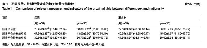
方法:收集于关节外科就诊的患者60例,其中汉族组、蒙古族组各30例,各组均男15例,女15例,年龄(36.00±7.22)岁。16排螺旋CT扫描仪(美国GE公司Lightspeed 16)对其进行螺旋扫描,层厚0.625 mm,扫描图像以DICOM格式导出存盘。再利用Mimics 15.0三维重建软件进行数字化三维重建测量。分别对胫骨平台宽、胫骨平台内侧前后径和外侧前后径进行测量,以观察其在性别、侧别和民族间是否具有差异性。
结果与结论:以上各指标值在左、右侧别间差异均无显著性意义(P > 0.05)。胫骨平台宽、胫骨平台内侧前后径和胫骨平台外侧前后径在男、女性别间和汉族、蒙古族之间差异均存在显著性意义(P < 0.05),具体表现在:①胫骨近端的形态学测量参数无论汉族组还是蒙古族组在性别间差异均存在显著性意义,且男性各均值大于女性。②部分参数指标在汉族与蒙古族间存在着一定的差异性,提示在施行人工全关节置换时,应根据国人形态特征、性别、民族、地区等差异正确地选择并放置假体。③数字化三维重建技术和个体化设计可以针对不同人群选择适合的假体,从而确保患者在全膝关节置换后获得良好的修复效果。
中图分类号:
.jpg)
.jpg)
.jpg)