| [1] 杨燕子.牙颌畸形患者正畸中自锁托槽矫治器对降低牙周指数、龈沟液AST含量的作用[J].中国伤残医学,2015,23(12):69-70.
[2] 董玉婉,赵秋颖,贾芳芳,等.纳米银羟基磷灰石涂层陶瓷托槽的研制及力学性能研究[J].口腔颌面修复学杂志,2014,15(3): 134-136.
[3] 董玉婉,赵秋颖,贾芳芳,等.纳米银羟基磷灰石涂层陶瓷托槽的抗菌性研究[J].中华老年口腔医学杂志,2013,11(6):330-332.
[4] 董玉婉.纳米银羟基磷灰石涂层陶瓷托槽的力学及抗菌性能研究[D].解放军医学院,2014.
[5] Ma'mani L,Sheykhan M,Heydari A,et al.Nanosilver embedded on hydroxyapatite-encapsulated γ-Fe2O3: Superparamagnetic catalyst for chemoselective oxidation of primary amines to N-monoalkylated hydroxylamines.Appl Catal A Gen.2011;395(1/2):34-38.
[6] 郑学斌,季珩,黄静琪,等.等离子喷涂抗菌羟基磷灰石涂层研究[J].无机材料学报,2006,21(3):764-768.
[7] 徐伏秋,陈华军,丁梧秀,等.载银羟基磷灰石抗菌粉体和陶瓷的制备及抗菌性能[J].无机化学学报,2013,29(12):2582-2586.
[8] 杨辉,蔡日强,王栋,等.抗菌羟基磷灰石的一步合成及其表征[J].功能材料,2010,41(3):406-409.
[9] 朱梓园,张富强,郑学斌,等.含银抗菌羟基磷灰石涂层银离子的缓释性能[J].上海口腔医学,2009,18(1):66-68.
[10] [李轩琦,孙康宁,卢志华,等.载银羟基磷灰石复合抗菌材料的研究[J].硅酸盐通报,2008,27(1):12-15,25.
[11] 师彩丽,苏炳煌,邱艳静,等.载银羟基磷灰石抗菌陶瓷粉体的应用研究[J].功能材料,2004,35(z1):2381-2382,2385.
[12] 陈有应,耿彬,王金清,等.含氟、锌、银羟基磷灰石涂层的制备与结构表征[J].材料导报,2012,26(z1):267-269.
[13] Saravanan S,Nethala S,Pattnaik S,et al.Preparation, characterization and antimicrobial activity of a bio-composite scaffold containing chitosan/nano-hydroxyapatite/nano-silver for bone tissue engineering.Int J Biol Macromol. 2011; 49(2): 188-193.
[14] 李箐,冯希平,廖运茂,等.钛表面载银HA-TCP溶胶凝胶涂层的制备及其抗茵性的研究[J].中国口腔颌面外科杂志,2008,6(2): 127-130.
[15] 朱梓园,张富强,郑学斌,等.钛种植体载银抗菌羟基磷灰石涂层的制备及结构特征[J].上海口腔医学,2006,15(5):543-546.
[16] 胡苏宁,张敏.钛表面载银硅羟基磷灰石镀膜对口腔常见菌抑菌作用的研究[C].//第七次全国口腔材料学术交流会论文集,2011: 73-74.
[17] 史建陆,魏婷婷,林奕真,等.微种植钉表面含银羟基磷灰石涂层的制备及溶血试验[J].临床口腔医学杂志,2012,28(6):336-339.
[18] 张敏,史建陆,林昌建,等.含银羟基磷灰石种植体的细胞毒性研究[J].临床口腔医学杂志,2009,25(3):139-141.
[19] Stanic V,Janackovic D,Dimitrijevic S,et al.Synthesis of antimicrobial monophase silver-doped hydroxyapatite nanopowders for bone tissue engineering.Appl Surf Sci.2011; 257(9):4510-4518.
[20] 彭伟伟,翟万银,江龙,等.人牙髓细胞复合不同孔径羟基磷灰石/磷酸三钙支架材料的生物学行为[J].中国组织工程研究与临床康复,2010,14(51):9567-9571.
[21] Oudadesse H,Mostafa A,Legal Y,et al.Elaboration and physicochemical behaviour of Nano Hydroxyapatite / Silver Nanoparticles biocompsite after 'in vitro' assays[C].//Recent advances in applied & biomedical informatics and computational engineering in systems applications.2011: 71-76.
[22] 王宇华,朱梓园,张富强,等.单质银应用于等离子喷涂抗菌涂层的研究[J].上海口腔医学,2009,18(3):317-319.
[23] Nirmala R,Nam KT,Park DK,et al.Structural, thermal, mechanical and bioactivity evaluation of silver-loaded bovine bone hydroxyapatite grafted poly(ε-caprolactone) nanofibers via electrospinning.Surf Coat Technol. 2010; 205(1):174-181.
[24] Nirmala R,Kanjwal MA,LeeJH,et al.Synthesis and characterization of bovine femur bone hydroxyapatite containing silver nanoparticles for the biomedical applications. J Nanopart Res.2011;13(5):1917-1927.
[25] 张敏,应志敏,史建陆,等.钛表面电化学沉积羟基磷灰石/纳米银涂层及体外细胞毒性评价[J].口腔颌面修复学杂志, 2010, 11 (5):312-315.
[26] 张敏,施更生,林海升,等.纳米银羟基磷灰石复合涂层的生物安全性研究[J].口腔医学,2010,30(9):541-543.
[27] 张敏,史建陆,林昌健,等.纳米银/羟基磷灰石涂层的制备与表征[J].中国口腔种植学杂志,2010,15(1):14-16,47.
[28] 陈梦云,陈素华,陈昆达,等.银离子羟基磷灰石糊剂治疗狗慢性根尖周炎的实验研究[J].口腔材料器械杂志,2006,15(1):47,50.
[29] 王晨,王朝俭,张焱,等.羟基磷灰石涂层种植体植入生物体内结合过程的动物实验观察[J].宁夏医学杂志,1999,21(6):341-342.
[30] 王臻,刘伟强.纳米复合树脂口腔修复材料的研制及性能表征[C].//第八届中国纳米科技西安研讨会论文集,2009:51-58.
[31] Nida Iqbal,Mohammed Rafiq Abdul Kadir,Nik Ahmad Nizam Nik Malek,et al.Rapid microwave assisted synthesis and characterization of nanosized silver-doped hydroxyapatite with antibacterial properties.Mater Lett.2012;89:118-122.
[32] 白丁,刘筱琳.Nd:YAG激光辅助去除陶瓷托槽对托槽的粘接强度及髓腔温度影响的研究[J].华西口腔医学杂志, 2004, 22(4): 287-289.
[33] Sheikh FA,Barakat NA,Kanjwal MA,et al.Electrospun titanium dioxide nanofibers containing hydroxyapatite and silver nanoparticles as future implant materials.J Mater Sci Mater Med.2010;21(9):2551-2559.
[34] Marsich E,Bellomo F,Turco G,et al.Nano-composite scaffolds for bone tissue engineering containing silver nanoparticles: preparation, characterization and biological properties.J Mater Sci Mater Med.2013;24(7):1799-1807.
[35] Pishbin F,Mouriño V,Gilchrist JB,et al.Single-step electrochemical deposition of antimicrobial orthopaedic coatings based on a bioactive glass/chitosan/nano-silver composite system.Acta Biomaterialia.2013;9(7):7469-7479.
[36] 仇玲玲,白玉兴,厉松,等.不同陶瓷托槽与金属弓丝间摩擦力的研究[J].北京口腔医学,2011,19(5):280-282.
[37] Koizhaiganova M,Lkhagvajav N,Ya?a I,et al.New gelatin-hydroxyapatite nanocomposite films containing nano silver: Synthesis, and, mechanical and antimicrobial properties. J Bionanosci.2011;5(2):130-137.
[38] 王卫东,董苁蓉,李蒙,等.陶瓷托槽与金属托槽对牙周指数的影响及其脱落率比较[J].广东牙病防治,2014,22(6):328-330.
[39] 王卫东,董苁蓉,李蒙,等.多晶氧化铝陶瓷托槽与金属托槽对牙周指数的影响及其脱落率的对比观察[J].临床口腔医学杂志, 2014, 30(6):374-375. |
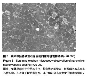
.jpg)
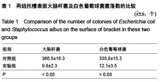
.jpg)
.jpg)