|
[1] Rai JJ,Kalantharakath T.Biomimetic ceramics for periodontal regeneration in infrabony defects: A systematic review.J Int Soc Prev Community Dent.2014;4(2):S78-S92.
[2] Ng VY.Risk of disease transmission with bone allograft. Orthopedics.2012; 35(8):679-681.
[3] Poinern GE,Brundavanam RK,Thi Le X,et al.The synthesis, characterisation and in vivo study of a bioceramic for potential tissue regeneration applications.Sci Rep.2014;4:6235.
[4] Berberi A,Samarani A,Nader N,et al.Physicochemical characteristics of bone substitutes used in oral surgery in comparison to autogenous bone. Biomed Res Int.2014; 2014: 320790.
[5] Albrektsson T,Johansson C.Osteoinduction, osteoconduction and osseointegration. Eur Spine J.2001;10(Suppl 2): S96-S101.
[6] Hannink G,Arts JJ.Bioresorbability, porosity and mechanical strength of bone substitutes: what is optimal for bone regeneration? Injury.2011;42 Suppl 2:S22-25.
[7] Barradas AM,Yuan H,van Blitterswijk CA,et al. Osteoinductive biomaterials: current knowledge of properties, experimental models and biological mechanisms.Eur Cell Mater. 2011;21: 407-429.
[8] LeGeros RZ. Calcium phosphate-based osteoinductive materials.Chem Rev.2008;108: 4742-4753.
[9] Karageorgiou V,Kaplan D.Porosity of 3D biomaterial scaffolds and osteogenesi. Biomaterials.2005;26(27):5474-5491.
[10] Gao C,Deng Y,Feng P,et al.Current Progress in Bioactive Ceramic Scaffolds for Bone Repair and Regeneration.Int J Mol Sci.2014;15(3):4714-4732.
[11] Tsuruga E,Takita H,Itoh H,et al.Pore size of porous hydroxyapatite as the cell-substratum controls BMP-induced osteogenesis.J Biochem.1997;121(2):317-324.
[12] Murphy CM,Haugh MG,O'Brien FJ.The effect of mean pore size on cell attachment, proliferation and migration in collagen-glycosaminoglycan scaffolds for bone tissue engineering.Biomaterials.2010;31(3):461-466.
[13] Jin QM,Takita H,Kohgo T,et al.Effects of geometry of hydroxyapatite as a cell substratum in BMP-induced ectopic bone formation.J Biomed Mater Res.2000;51:491-499.
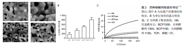
[14] Yuan H,Fernandes H,Habibovic P,et al.Osteoinductive ceramics as a synthetic alternative to autologous bone grafting.Proc Natl Acad Sci USA.2010;107(31): 13614-13619.
[15] Oh SH,Kim TH,Im GI,et al.Investigation of pore size effect on chondrogenic differentiation of adipose stem cells using a pore size gradient scaffold. Biomacromolecules. 2010;11(8): 1948-1955.
[16] Hing KA. Bone repair in the twenty-first century: biology, chemistry or engineering?Philos Trans A Math Phys Eng Sci. 2004 ;362(1825):2821-2850.
[17] Lane JM ,Bostrom MP.Bone grafting and new composite biosynthetic graft materials. Instr Course Lect.1998;47: 525-534.
[18] Zhang X,Chang W,Lee P,et al.Polymer-ceramic spiral structured scaffolds for bone tissue engineering:effect of hydroxyapatite composition on human fetal osteoblasts.PLoS One.2014;9(1):e85871.
[19] Meng J,Xiao B,Zhang Y,et al.Super-paramagnetic responsive nanofibrous scaffolds under static magnetic field enhance osteogenesis for bone repair in vivo.Sci Rep. 2013; 3:2655.
[20] Sergey V,Dorozhkin.Biocomposites and hybrid biomaterials based on calcium orthophosphates.Biomatter. 2011;1(1): 3-56.
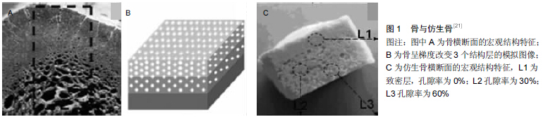
[21] Wang Q,Wang Q,Wan C.Preparation and evaluation of a biomimetic scaffold with porosity gradients in vitro.An Acad Bras Cienc.2012;84(1):9-16.
[22] Lindner M,Bergmann C,Telle R,et al.Calcium phosphate scaffolds mimicking the gradient architecture of native long bones.J Biomed Mater Res A.2014;102(10):3677-3684.
[23] Tampieri A,Celotti G,Sprio S,et al.Porosity-graded hydroxyapatite ceramics to replace natural bone.Biomaterials. 2001;22:1365-1370.
[24] Motomiya M,Ito M,Takahata M,et al.Effect of Hydroxyapatite porous characteristics on healing outcomes in rabbit posterolateral spinal fusion model.Eur Spine J.2007;16(12): 2215-2224.
[25] Teraoka K,Kato T,Hattori K,et al.Evaluation of the capacity of mosaic-like porous ceramics with designed pores to support osteoconduction.J Biomed Mater Res A. 2013;101(12): 3571-3579.
[26] Xiao X,Wang W,Liu D,et al.The promotion of angiogenesis induced by three-dimensional porous beta-tricalcium phosphate scaffold with different interconnection sizes via activation of PI3K/Akt pathways.Sci Rep.2015;5:9409.
[27] Wang L,Zhang B,Bao C,et al.Ectopic Osteoid and Bone Formation by Three Calcium-Phosphate Ceramics in Rats, Rabbits and Dogs.PLoS One.2014;9(9):e107044.
[28] Kandori K,Horigami N,Kobayashi H,et al.Adsorption of lysozome onto various synthetic hydroxyapatites.J Colloid Interface Sci. 1997;191:498-502.
[29] Uludag H, D’Augusta D,Palmer R,et al. Characterization of RHBMP-2 pharmacokinetics implanted with biomaterial carriers in the rat ectopic model.J Biomed Mater Res.1999;46: 193-202.
[30] Ito-Amano M,Nakamura Y,Morisaki M,et al.Temporal and Spatial Expression Patterns of Bone Morphogenetic Protein 3 in Developing Zebrafish.Open Rheumatol J.2014;8:69-72.
[31] Cui Q,Dighe AS,Irvine JN Jr.Combined angiogenic and osteogenic factor delivery for bone regenerative engineering. Curr Pharm Des.2013;19(19):3374-3383.
[32] Velasco MA,Narváez-Tovar CA,Garzón-Alvarado DA,et al. Design, Materials, and Mechanobiology of Biodegradable Scaffolds for Bone Tissue Engineering.Biomed Res Int. 2015; 2015:729076.
[33] Fauzi Kamal A,Hadisoebroto Dilogo I,Untung Hutagalung E,et al.Transplantation of mesenchymal stem cells, recombinant human BMP-2,and their combination in accelerating the union after osteotomy and increasing, the mechanical strength of extracorporeally irradiated femoral autograft in rat models.Med J Islam Repub Iran.2014;28:129.
[34] Bosemark P,Perdikouri C,Pelkonen M,et al.The masquelet induced membrane technique with BMP and a synthetic scaffold can heal a rat femoral critical size defect.J Orthop Res.2015; 33(4):488-495.
[35] Machado EG,Issa JP,Figueiredo FA,et al.A new heterologous fibrin sealant as scaffold to recombinant human bone morphogenetic protein-2 (rhBMP-2) and natural latex proteins for the repair of tibial bone defects.Acta Histochem.2015; 117(3): 288-296.
[36] Kim IG, Hwang MP, Du P, et al.Bioactive cell-derived matrices combined with polymer mesh scaffold for osteogenesis and bone healing.Biomaterials.2015;50:75-86.
[37] Ren X, Bischoff D, Weisgerber DW,et al.Osteogenesis on nanoparticulate mineralized collagen scaffolds via autogenous activation of the canonical BMP receptor signaling pathway.Biomaterials.2015;50:107-114.
[38] Eap S,Keller L,Schiavi J,et al.A living thick nanofibrous implant bifunctionalized with active growth factor and stem cells for bone regeneration.Int J Nanomedicine.2015;10: 1061-1075.
[39] Yang HL,Sun H.Calcium Phosphate Scaffolds Combined with Bone Morphogenetic Proteins or Mesenchymal Stem Cells in Bone Tissue Engineering.Chin Med J (Engl). 2015;128(8): 1121-1127.
[40] Ripamonti U, Crooks J, Kirkbride AN .Sintered porous hydroxyapatites with intrinsic osteoinductive activity: Geometric induction of bone formation.South African J Sci. 1999;95: 335-343.
[41] De Groot J.Carriers that concentrate native bone morphogenetic protein in vivo. Tissue Eng.1998;4: 337-341.
[42] Ripamonti U,Crooks J,Khoali L,et al.The induction of bone formation by coral-derived calcium carbonate/hydroxyapatite constructs.Biomaterials.2009;30: 1428-1439.
[43] Gallardo-López A,Domínguez-Rodríguez A,Estournès C,et al.Plastic deformation of dense nanocrystalline yttrium oxide at elevated temperatures.J Eur Ceram Soc. 2012;32: 3115-3121.
[44] Mukhopadhyay A,Basu B.Consolidation–microstructure– property relationships in bulk nanoceramics and ceramic nanocomposites: a review.Int Mater Rev.2007;52(Issue 5): 257-288.
[45] Dalby MJ,Gadegaard N,Tare R,et al.The control of human mesenchymal cell differentiation using nanoscale symmetry and disorder.Nat Mater.2007;6(12):997-1003.
[46] Despang F,Bernhardt A,Lode A,et al.Synthesis and physicochemical, in vitro and in vivo evaluation of an anisotropic, nanocrystalline hydroxyapatite bisque scaffold with parallel-aligned pores mimicking the microstructure of cortical bone.J Tissue Eng Regen Med.2013.doi: 10.1002/term.1729. [Epub ahead of print]
[47] Puzari A,Borah JP.Ionic self-assembly and hierarchies of polymeric structures generating nanoscale architecture: Opportunities ahead from industrial perspective. Rev Adv Mater Sci.2013;34:88-106.
[48] Chun YW, Crowder SW, Mehl SC,et al.Therapeutic application of nanotechnology in cardiovascular and pulmonary regeneration. Comput Struct Biotechnol J. 2013;7: e201304005.
[49] Feng P,Niu M,Gao C,et al. A novel two-step sintering for nano-hydroxyapatite scaffolds for bone tissue engineering.Sci Rep.2014;4:5599.
[50] Isobe T,Ooyama A,Shimizu M,et al.Pore size control of Al2O3 ceramics using two-step sintering.Ceram Int.2012;38: 787-793.
[51] Järvenpää A,Mäntyjärvi K,Merklein M,et al.Mechanical properties of laser heat treated 6 mm thick UHSS-steel.AIP Conf Proc.2011;1353:1319.
[52] Zhang J,Luo X,Barbieri D,et al.The size of surface microstructures as an osteogenic factor in calcium phosphate ceramics.Acta Biomater.2014;10(7):3254-3263.
[53] Lv R,Zhou W,Shi K,et al.Alumina decorated TiO nanotubes with ordered mesoporous walls as high sensitivity of NO gas sensors at room temperature.Nanoscale. 2013;5(18): 8569-8576.
[54] Javed F,Vohra F,Zafar S,et al.Significance of osteogenic surface coatings on implants to enhance osseointegration under osteoporotic-like conditions.Implant Dent. 2014;23(6): 679-86.
[55] Motamedian SR,Hosseinpour S,Ahsaie MG,et al.Smart scaffolds in bone tissue engineering: A systematic review of literature.World J Stem Cells.2015;7(3):657-668.
[56] Duan Y,Lü W,Wang C,et al.The effects of surface morphology of calcium phosphate ceramics on apatite formation in dynamic SBF.Sheng Wu Yi Xue Gong Cheng Xue Za Zhi. 2002;19:186-190.
[57] Refai AK,Textor M,Brunette DM,et al.Effect of titanium surface topography on macrophage activation and secretion of proinflammatory cytokines and chemokines. J Biomed Mater Res A.2004;70:194-205.
[58] Chen S,Jones JA,Xu Y,et al.Characterization of Topographical Effects on Macrophage Behavior in a Foreign Body Response Model.Biomaterials.2010;31(13):3479-3491.
[59] Klar RM,Duarte RDix-Peek T,et al.Calcium ions and osteoclastogenesis initiate the induction of bone formation by coral-derived macroporous constructs.Cell Mol Med.2013; 17(11):1444-1457.
[60] Zhang J,Barbieri D,ten Hoopen H,et al.Microporous calcium phosphate ceramics driving osteogenesis through surface architecture.J Biomed Mater Res A. 2015;103(3):1188-1199. |