| [1] Reid IR. Should we prescribe calcium supplements for osteoporosis prevention. J Bone Metab. 2014;21(1):21-28.
[2] de Waure C, Specchia ML, Cadeddu C, et al. The prevention of postmenopausal osteoporotic fractures: results of the Health Technology Assessment of a new antiosteoporotic drug. Biomed Res Int. 2014;2014:975927.
[3] Levine JP. Pharmacologic and nonpharmacologic management of osteoporosis. Clin Cornerstone. 2006;8(1):40-53.
[4] Sanders S, Geraci SA. Osteoporosis in postmenopausal women: considerations in prevention and treatment: (women's health series). South Med J. 2013;106(12): 698-706.
[5] Inada M, Matsumoto C, Miyaura C. Animal models for bone and joint disease. Ovariectomized and orchidectomized animals. Clin Calcium. 2011;21(2):164-170.
[6] Nakamuta H. The ovariectomized animal model of postmenopausal bone loss. Nihon Rinsho. 2004;62 Suppl 2:759-763.
[7] Clément-Lacroix P, Ai M, Morvan F, et al. Lrp5-independent activation of Wnt signaling by lithium chloride increases bone formation and bone mass in mice. Proc Natl Acad Sci U S A. 2005;102(48):17406-17411.
[8] 高丽娜,安莹,杨昊,等.氯化锂对人颌骨来源的骨髓间充质干细胞增殖及骨向分化能力的影响[J].实用口腔医学杂志,2013,29(2): 203-208.
[9] Smith G, Stassen L, Flint S. Treating osteoporosis. Ir Med J. 2009;102(3):88.
[10] Cooper C, Harvey N, Cole Z, et al. Developmental origins of osteoporosis: the role of maternal nutrition. Adv Exp Med Biol. 2009;646:31-39.
[11] 杨玺.骨质疏松症合理用药[M]. 北京:科学技术文献出版社, 2008:104-105.
[12] Miao Q, Li JG, Miao S, et al. The bone-protective effect of genistein in the animal model of bilateral ovariectomy: roles of phytoestrogens and PTH/PTHR1 against post-menopausal osteoporosis. Int J Mol Sci. 2012;13(1):56-70.
[13] Taipale J, Beachy PA. The Hedgehog and Wnt signalling pathways in cancer. Nature. 2001;411(6835):349-354.
[14] Peifer M, Polakis P. Wnt signaling in oncogenesis and embryogenesis--a look outside the nucleus. Science. 2000; 287(5458):1606-1609.
[15] Hartmann C. A Wnt canon orchestrating osteoblastogenesis. Trends Cell Biol. 2006;16(3):151-158.
[16] Monroe DG, McGee-Lawrence ME, Oursler MJ, et al. Update on Wnt signaling in bone cell biology and bone disease. Gene. 2012;492(1):1-18.
[17] Westendorf JJ, Kahler RA, Schroeder TM. Wnt signaling in osteoblasts and bone diseases. Gene. 2004;341:19-39.
[18] Kikuchi A. Regulation of beta-catenin signaling in the Wnt pathway. Biochem Biophys Res Commun. 2000;268(2): 243-248.
[19] Kim HS, Skurk C, Thomas SR, et al. Regulation of angiogenesis by glycogen synthase kinase-3beta. J Biol Chem. 2002;277(44):41888-41896.
[20] Hu H, Hilton MJ, Tu X, et al. Sequential roles of Hedgehog and Wnt signaling in osteoblast development. Development. 2005;132(1):49-60.
[21] Bennett CN, Longo KA, Wright WS, et al. Regulation of osteoblastogenesis and bone mass by Wnt10b. Proc Natl Acad Sci U S A. 2005;102(9):3324-3329.
[22] Luo Y, Cai J, Xue H, et al. SDF1alpha/CXCR4 signaling stimulates beta-catenin transcriptional activity in rat neural progenitors. Neurosci Lett. 2006;398(3):291-295.
[23] Jope RS. Lithium and GSK-3: one inhibitor, two inhibitory actions, multiple outcomes. Trends Pharmacol Sci. 2003; 24(9): 441-443.
[24] Danielsen CC, Mosekilde L, Svenstrup B. Cortical bone mass, composition, and mechanical properties in female rats in relation to age, long-term ovariectomy, and estrogen substitution. Calcif Tissue Int. 1993;52(1):26-33.
[25] Xiang A, Kanematsu M, Mitamura M, et al. Analysis of change patterns of microcomputed tomography 3-dimensional bone parameters as a high-throughput tool to evaluate antiosteoporotic effects of agents at an early stage of ovariectomy-induced osteoporosis in mice. Invest Radiol. 2006;41(9):704-712.
[26] 牛银波,李宇华,伍焕杰,等.氯化锂对去卵巢大鼠骨密度的影响研究[J]. 解放军医学杂志,2010, 35(1):54-57.
[27] Pino AM, Rosen CJ, Rodríguez JP. In osteoporosis, differentiation of mesenchymal stem cells (MSCs) improves bone marrow adipogenesis. Biol Res. 2012;45(3):279-287.
[28] Ducy P, Zhang R, Geoffroy V, et al. Osf2/Cbfa1: a transcriptional activator of osteoblast differentiation. Cell. 1997;89(5):747-754.
[29] Nakashima K, Zhou X, Kunkel G, et al. The novel zinc finger-containing transcription factor osterix is required for osteoblast differentiation and bone formation. Cell. 2002; 108(1):17-29.
[30] Pino AM, Rosen CJ, Rodríguez JP. In osteoporosis, differentiation of mesenchymal stem cells (MSCs) improves bone marrow adipogenesis. Biol Res. 2012;45(3):279-287.
[31] Khan E, Abu-Amer Y. Activation of peroxisome proliferator-activated receptor-gamma inhibits differentiation of preosteoblasts. J Lab Clin Med. 2003;142(1):29-34. |
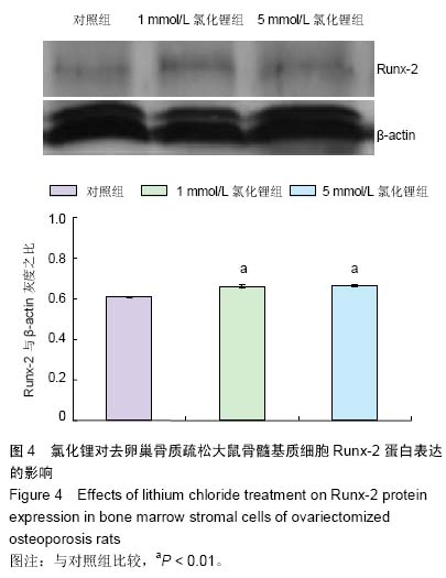
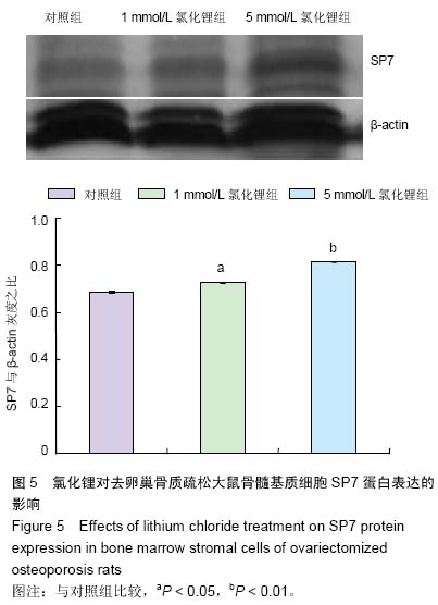
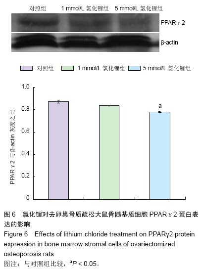
lb.jpg)