中国组织工程研究 ›› 2015, Vol. 19 ›› Issue (38): 6179-6183.doi: 10.3969/j.issn.2095-4344.2015.38.020

• 纳米生物材料 nanobiomaterials • 上一篇    下一篇

5-氟尿嘧啶聚乳酸载药纳米微粒对胃癌细胞株的体外杀伤作用

李晓利1,牛 敏1,张 铭1,张娜娜1,施 瑶2   

  1. 1西安市红会医院消化内科,陕西省西安市 710054; 
    2西安交通大学医学院第一附属医院消化内科,陕西省西安市 710061
  • 通讯作者: 李晓利,西安市红会医院消化内科,陕西省西安市 710054
  • 作者简介:李晓利,女,汉族、1966年生,陕西省人,1990年西安交通大学医学院毕业,副主任医师。
  • 基金资助:

      西安交通大学医学院第一附属医院科研基金(2014YK.5)

5-Fluorouracil-loaded polylactic acid nanoparticles have a killing effect on gastric cancer cell lines in vitro

Li Xiao-li1, Niu Min1, Zhang Ming1, Zhang Na-na1, Shi Yao2   

  1. 1Department of Gastroenterology, Xi’an Honghui Hospital, Xi’an 710054, Shaanxi Province, China;
     2Department of Gastroenterology, the First Affiliated Hospital of Medical College of Xi’an Jiaotong University, Xi’an 710061, Shaanxi Province, China
  • Contact: Li Xiao-li, Department of Gastroenterology, Xi’an Honghui Hospital, Xi’an 710054, Shaanxi Province, China
  • About author:Li Xiao-li, Associate chief physician, Department of Gastroenterology, Xi’an Honghui Hospital, Xi’an 710054, Shaanxi Province, China
  • Supported by:

    the Scientific Research Foundation of the First Affiliated Hospital of Medical College of Xi’an Jiaotong University, No. 2014YK.5

摘要:

背景:5-氟尿嘧啶在胃癌治疗中占据重要地位,但是长期服用容易出现骨髓抑制、白细胞减少等不良反应。聚乳酸及共聚物载药微粒材料生物相容性较高,分解物不会在机体内发生聚集。

目的:探讨聚乳酸载药纳米微粒对胃癌细胞株的体外杀伤作用机制。
方法:选取10只小鼠进行实验,利用超声乳化方法制备5-氟尿嘧啶聚乳酸载药纳米微粒,并采用噻唑蓝比色法配制1×10-7,1×10-6,1×10-5,1×10-4 mol/L的5-氟尿嘧啶聚乳酸载药纳米微粒,检测其对小鼠胃癌细胞的体外杀伤效应,并且计算出药物的抑制率浓度以及对胃癌细胞的生长抑制能力等以及凋亡的诱导作用。
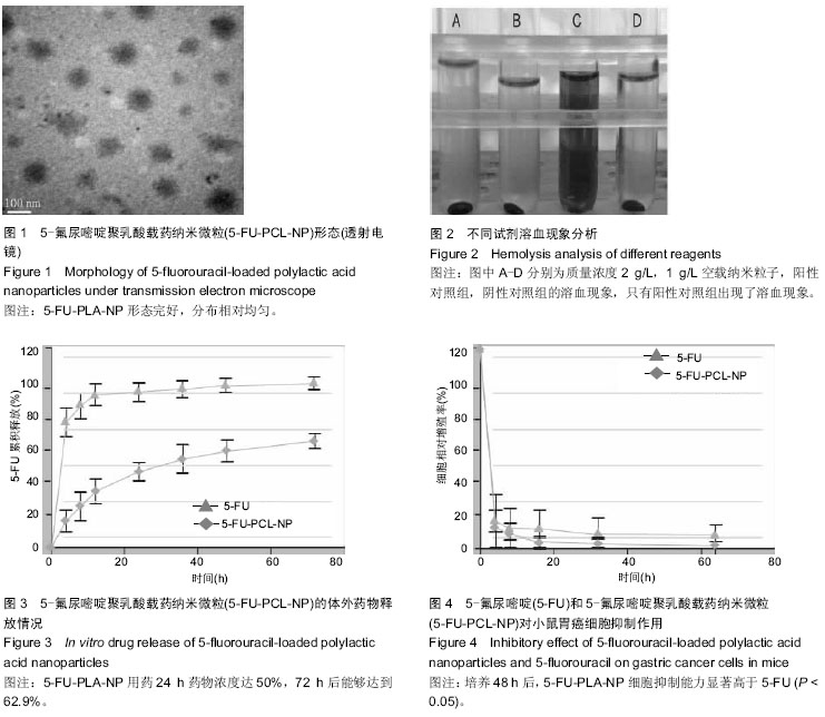
结果与结论:透射电镜下观察5-氟尿嘧啶聚乳酸载药纳米微粒:形态完好,分布相对均匀,不出现粘连等现象,用药24 h药物浓度可达到50%,72 h后能够达到62.9%;不同浓度下单一5-氟尿嘧啶和5-氟尿嘧啶聚乳酸载药纳米微粒和小鼠胃癌细胞联合培养48 h后,细胞的活性随着药物浓度的提高出现下降趋势,并且5-氟尿嘧啶聚乳酸载药纳米微粒细胞抑制能力显著高于5-氟尿嘧啶(P < 0.05);5-氟尿嘧啶聚乳酸载药纳米微粒的IC50显著低于5-氟尿嘧啶(P < 0.05)。结果证实聚乳酸纳米微粒具有优良的药物载体作用,载药量较大,在机体内提高药物浓度,并且不降低5-氟尿嘧啶成分的生物学活性,可为胃癌治疗提供新思路。

中国组织工程研究杂志出版内容重点:生物材料;骨生物材料; 口腔生物材料; 纳米材料; 缓释材料; 材料相容性;组织工程

关键词: 生物材料, 纳米材料, 药物控释材料, 聚乳酸载药纳米微粒, 小鼠胃癌细胞株, 体外杀伤, 5-氟尿嘧啶, 体外杀伤, 抑制率浓度, 生长抑制能力, 诱导作用, 生物半衰期

Abstract:

BACKGROUND: 5-Fluorouracil occupies an important position in the treatment of gastric cancer, but its long-term use can easily induce adverse reactions such as myelosuppression and leukopenia. Polylactic acid and its copolymers have a higher biocompatibility, and their decomposer cannot gather in the body.

OBJECTIVE: To investigate the in vitro cytotoxicity mechanism of 5-fluorouracil-loaded polylactic acid nanoparticles on gastric cancer cell lines.
METHODS: Ten mice were selected in this study. 5-fluorouracil-loaded polylactic acid nanoparticles (1×10⁻⁷, 1×10⁻⁶, 1×10⁻⁵, 1×10⁻⁴ mol/L) were prepared using ultrasonic emulsification method. Killing effect of polylactic acid nanoparticles on gastric cancer cell lines in vitro were detected. Then, the inhibition rate was calculated at different concentrations.
RESULTS AND CONCLUSIONS: Under the transmission electron microscope, 5-fluorouracil-loaded polylactic acid nanoparticles had good shape and relatively evenly distributed with no adhesions. After drug administration, the drug concentration was 50% at 24 hours and 62.9% at 72 hours. After 48 hours co-culture with single 5-fluorouracil or 5-fluorouracil-loaded polylactic acid nanoparticles, the viability of gastric cancer cells showed a decrease trend with the increase of drug concentrations, and moreover, 5-fluorouracil-loaded polylactic acid nanoparticles had a better cell inhibition ability than the single 5-fluorouracil (P < 0.05). The IC50 value of 5-fluorouracil-loaded polylactic acid nanoparticles was significantly lower than that of 5-fluorouracil (P < 0.05). These findings indicate that polylactic acid nanoparticles as good drug carriers have a strong drug loading capacity and increase drug concentration in the body, but cannot reduce the biological activity of 5-fluorouracil, which provide new ideas for the treatment of gastric cancer.

中国组织工程研究杂志出版内容重点:生物材料;骨生物材料; 口腔生物材料; 纳米材料; 缓释材料; 材料相容性;组织工程

Key words: Tissue Engineering, Biocompatible Materials, Stomach Neoplasms

中图分类号: