中国组织工程研究 ›› 2015, Vol. 19 ›› Issue (16): 2534-2539.doi: 10.3969/j.issn.2095-4344.2015.16.014

• 材料生物相容性 material biocompatibility • 上一篇    下一篇

纳米羟基磷灰石对人脐带静脉血管内皮细胞的毒性评价

程光存,严中亚,李春生,严  宇,韦晓勇   

  1. 安徽医科大学附属省立医院胸心外科,安徽省心血管病研究所,安徽省合肥市  230001
  • 收稿日期:2015-03-01 出版日期:2015-04-16 发布日期:2015-04-16
  • 通讯作者: 严中亚,教授,主任医师,安徽医科大学附属省立医院胸心外科,安徽省心血管病研究所,安徽省合肥市 230001
  • 作者简介:程光存,男,1966年生,安徽省舒城市人,硕士,副主任医师,副教授,主要从事体外循环在在心脏外科学中的应用研究。
  • 基金资助:

    安徽省自然科学基金项目(1308085MH143)

Nano-hydroxyapatite is non-toxic to human umbilical cord vein endothelial cells

Cheng Guang-cun, Yan Zhong-ya, Li Chun-sheng, Yan Yu, Wei Xiao-yong   

  1. Department of Cardiothoracic Surgery, the Affiliated Provincial Hospital of Anhui Medical University, Anhui Provincial Institute of Cardiovascular Disease, Hefei 230001, Anhui Province, China
  • Received:2015-03-01 Online:2015-04-16 Published:2015-04-16
  • Contact: Yan Zhong-ya, Professor, Chief physician, Department of Cardiothoracic Surgery, the Affiliated Provincial Hospital of Anhui Medical University, Anhui Provincial Institute of Cardiovascular Disease, Hefei 230001, Anhui Province, China
  • About author:Cheng Guang-cun, Master, Associate chief physician, Associate professor, Department of Cardiothoracic Surgery, the Affiliated Provincial Hospital of Anhui Medical University, Anhui Provincial Institute of Cardiovascular Disease, Hefei 230001, Anhui Province, China
  • Supported by:

    the Natural Science Foundation of Anhui Province, No. 1308085MH143

摘要:

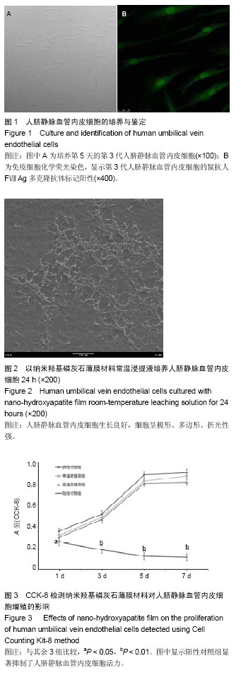
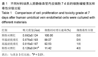
背景:有研究应用脉冲激光沉积合成技术在人工心脏机械瓣膜上沉积胶原制备了新型纳米羟基磷灰石薄膜涂层。
目的:观察此种新型纳米羟基磷灰石薄膜对人脐带静脉血管内皮细胞的毒性。
方法:分别采用纳米羟基磷灰石薄膜常温浸提液、纳米羟基磷灰石薄膜高温浸提液、高密度聚乙烯及苯酚溶液培养人脐静脉血管内皮细胞,72 h内倒置相差显微镜下观察细胞生长状况;培养7 d时采用CCK-8法检测细胞增殖与毒性分级。
结果与结论:培养24 h,纳米羟基磷灰石薄膜常温浸提液组、纳米羟基磷灰石薄膜高温浸提液组和高密度聚乙烯组细胞生长良好,呈梭形,折光性强,3组细胞形态、数量无明显差异;苯酚溶液组细胞多为悬浮、圆形、固缩的死细胞;48 h时,除了苯酚溶液组外,其余3组细胞数量明显增加,细胞生长密集,至72 h时细胞生长旺盛,间隙显著减小。培养7 d内,纳米羟基磷灰石薄膜常温浸提液组、纳米羟基磷灰石薄膜高温浸提液组和高密度聚乙烯组细胞增殖活性无差异,均高于苯酚溶液组(P < 0.05),纳米羟基磷灰石薄膜毒性级别为0至1级,表明纳米羟基磷灰石人工心脏机械瓣膜有良好的组织细胞相容性,无毒性作用。


中国组织工程研究杂志出版内容重点:生物材料;骨生物材料; 口腔生物材料; 纳米材料; 缓释材料; 材料相容性;组织工程


全文链接:

关键词: 生物材料, 材料相容性, 羟基磷灰石, 人脐带静脉血管内皮细胞, 细胞毒性, 活细胞计数试剂盒, 安徽省自然科学基金

Abstract:

BACKGROUND: Pulsed laser deposition synthesis technology has been used to prepare new nano-hydroxyapatite thin film coating by collagen deposition on artificial mechanical heart valve. 
OBJECTIVE: To investigate the toxicity of new nano-hydroxyapatite thin film on human umbilical vein endothelial cells.
METHODS: Human umbilical vein endothelial cells were cultured with nano-hydroxyapatite film room-temperature leaching solution, nano-hydroxyapatite film high-temperature leaching solution, high-density polyethylene and phenol solution. Within 72 hours, cell growth was observed under the inverted phase contrast microscope. At 7 days after culture, cell proliferation and toxicity grading were detected using Cell Counting Kit-8. 
RESULTS AND CONCLUSION: At 24 hours after culture, cells grew well, showed fusiform shape, and had strong refraction in the nano-hydroxyapatite film room-temperature leaching solution, nano-hydroxyapatite film high-temperature leaching solution, high-density polyethylene groups, and no significant differences in cell morphology and number were detected among above groups. Cells in the phenol solution group were suspended, round, pyknotic and dead. At 48 hours, except phenol solution group, cell number increased significantly, and cell grew densely in other three groups. At 72 hours, cells grew strongly, and the gap became small obviously. Within 7 days after culture, cell proliferation activity was not significant in the nano-hydroxyapatite film room-temperature leaching solution, nano-hydroxyapatite film high-temperature leaching solution, and high-density polyethylene groups, which was significantly higher than in the phenol solution group (P < 0.05). The toxicity of nano-hydroxyapatite film graded 0 to 1. These results suggested that nano-hydroxyapatite artificial mechanical heart valve has good histocompatibility, but no toxicity.


中国组织工程研究杂志出版内容重点:生物材料;骨生物材料; 口腔生物材料; 纳米材料; 缓释材料; 材料相容性;组织工程


全文链接:

Key words: Durapatite, Endothelial Cells, Umbilical Veins

中图分类号: