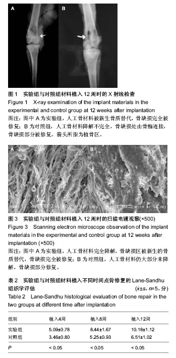
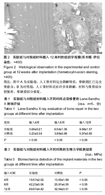
| [1] Kikuchi M,Itoh S,Ichinose S,et al.Self-organization mechanism in a bone-like hydroxyapatite/collagen nanocomposite synthesized in vitro and its biological reaction in vivo.Biomaterials.2001;22(13):1705-1711.
[2] 王小红,马建标,王亦农,等.骨修复材料的研究进展[J].生物医学工程学杂志,2001,18(4):647-652.
[3] 冯庆玲,崔福斋,张伟.纳米羟基磷灰石/胶原骨修复材料[J].中国医学科学院报,2002,24(2):124-128.
[4] Arosarena OA,Collins WL.Bone regeneration in the rat mandible with bone morphogenetic protein-2: a comparison of two carriers.Otolaryngol Head Neck Surg. 2005;132(4): 5922-5971.
[5] Deng X,Hao J,Wang C.Preparation and mechanical properties of nanocomposites of poly(D,L-lactide) with Ca-deficient hydroxyapatite nanocrystals. Biomaterials.2001; 22(21):2867-2873.
[6] Wang A.Effect of Head Cuponthewear of UHMWPE in Total Hip Replacement.Biomaterials.1998;Aprial 22-26:357.
[7] Goshima J,Goldberg VM,Caplan AI.Osteogenic potential of culture-expanded rat marrow cells as assayed in vivo with porous calcium phosphate ceramic.Biomterials.1991; 12(2): 253-258.
[8] Liao SS,Guan K,Cui FZ,et al.Lumbar spine fusion with a mineralized collagen matrix and rhBMP-2 in rabbit model.Spine (Phila Pa 1976).2003;28(17):1954-1960.
[9] Sun TS,Guan K,Shi SS,et al.Effect of nano-hydroxyapatite/ collagen composite and bone morphogenetic protein-2 on lumbar in transverse fusion in rabbits.Chin J Traumatol. 2004;7(1):18-24.
[10] Du C,Cui FZ,Zhang W,et al.Formation of calcium phosphate/collagen composited through mineralization of collagen matrix.J Biomed Mater Res.2000;50(4):518-527.
[11] Hartgerink JD,Beniash E,Stupp SI.Self-assembly and mineralization of peptide-amphiphile nanofibers.Science. 2001;294(5547):1684-1688.
[12] Yoshikawa M, Toda T.Reconstruction of alveolar bone defect by calcium phosphate compounds.J Biomed Mater Res. 2000;53(4): 430-437.
[13] Lang H,Mertens T,Gerlack KL.Re-implantation of homologous, cultivated osteoblast-like cells for improvement of bone regeneration. An animal study.Int Oral Maxi Surg.1989;18(4): 244-248.
[14] Engelberg I, Kohn J.Physico-mechanical properties of degradable polymers used in medical applications: a comparative study.Biomaterials.1991;12(3):292-304.
[15] Liu Q,de Wijin JR,van Blitterswijk CA.Nano-apatite/polymer composites: mechanical and physicochemical characteristics. Biomaterials.1997;18(9):1263-1270.
[16] Bagambisa FB,Kappert HF,Schilli W. Interfacial reactions of osteoblasts to dental and implant materials.J Oral Maxillofac Surg.1994;52(1):52-56.
[17] Williams DF.Titanium and titanium alloys.Biocompatibility of Implant Materials,CRC Press,Boca Raton,1981.
[18] 魏建华.活性复合钛基种植体表面的构建及生物学特性研究[D].解放军第四军医大学,2003.
[19] Hench LL,Splinter RJ,Allen WC,et al.Bonding mechanisms at the interface of ceramic prosthetic materials. J Biomed Mater Res.1972;2:117-141.
[20] Domer-Reisel A,Klenn V,Irmer G,et al.Nano- and microstructure of short fibre reinforced and unreinforced hydroxyaptite.Biomed Tech(Berl).2002;47 suppl 1 Pt 1: 397-400.
[21] 王学江,李玉宝.羟基磷灰石纳米针结晶与聚酰胺仿生复合生物材料研究[J].高技术通讯,2001,11(5):1-5.
[22] Bonfield W,Grynpas MD,Tully AE,et al.Hydroxyapatite reinforced polyethylene--a mechanically compatible implant material for bone replacement. Biomaterials.1981;2(3): 185-186.
[23] Karch J,Birringer R,Gleiter H.Ceramics ductile at low temperature.Nature.1987;330(6148):556-558.
[24] Sleytr UB,Pum D,Sára M.Advances in S-layer nanotechnology and biomimetics.Adv Biophys.1997;34:71-79.
[25] 魏红,李永国.纳米技术在生物医学工程领域的应用-研究现状和发展趋势[J].国外医学:生物医学工程分册,1999,22(6):340-345.
[26] Murphy J,Carr B,Atkinson T. Nanotechnology in medicing and the biosciences. Trends Biotechnol.1994;12(2):289-290.
[27] 骆小平,赵定风,田杰谟,等.华西医科大学学报,1998,29(24): 383-386.
[28] Webster TJ,Ergun C,Doremys RH,et al.Enhanced osteoclast- like cell function on nanophase ceramics. Biomaterials. 2001; 2(11):1327-1333.
[29] Cuneyt TA.Nanospace theory for biomineralization Biomaterials. 2000;21(1):1429-1438.
[30] Tas AC.Synthesis of biomimetic Ca-hydroxyapatite powders at 37 degrees C in synthetic body fluids.Biomaterials.2000; 21(14):1429-1438.
[31] Seeman NC.DNA nanotechnology: novel DNA constructions. Annu Rev Biophys Biomol Struct.1998;27: 225-248.
[32] Bouyer E,Gitzhofer F,Boulos MI.Morphological study of hydroxyapatite nanocrystal suspension.J Mater Sci Mater Med. 2000;11(8):523-531.
[33] Weiner S,Price PA.Disaggregation of bone into crystals.Calcif Tissue Int. 1986;39(6):365-375.
[34] Barralet JE, Gaunt T, Wright AJ, et al. Effect of porosity reduction by compaction on compressive strength and microstructure of calcium phosphate cement.J Biomed Mater Res.2002;63(1):1-9. |