中国组织工程研究 ›› 2014, Vol. 18 ›› Issue (23): 3691-3695.doi: 10.3969/j.issn.2095-4344.2014.23.014

• 干细胞培养与分化 stem cell culture and differentiation • 上一篇    下一篇

山羊神经干细胞的分离培养及体外诱导分化

夏许可,张 伟,余杰锋,章 莹   

  1. 1解放军广州军区广州总医院骨科医院,广东省广州市 510010;2南方医科大学研究生院 广东省广州市 510515;3广东冠昊生物科技股份有限公司,广东省广州市 510530;4广州医科大学研究生院,广东省广州市 510182
  • 修回日期:2014-04-17 出版日期:2014-06-04 发布日期:2014-06-04
  • 通讯作者: 章莹,博士,主任医师,解放军广州军区广州总医院骨科医院,广东省广州市 510010
  • 作者简介:夏许可,男,1987年生,北京市人,汉族,南方医科大学在读硕士,主要从事外周神经损伤的研究。
  • 基金资助:

    广东省科技计划项目(2010B031100017);广州市科技计划项目(2012J4100039)

In vitro culture and differentiationof goat neural stem cells

Xia Xu-ke1, 2, Zhang Wei3, Yu Jie-feng1, 4, Zhang Ying1   

  1. 1Department of Orthopedics, Guangzhou General Hospital of Guangzhou Military Command, Guangzhou 510010, Guangdong Province, China; 2Graduate School of Southern Medical University, Guangzhou 510515, Guangdong Province, China; 3Grandhope Biotech Co., Ltd., Guangzhou 510530, Guangdong Province, China; 4Graduate School of Guangzhou Medical University, Guangzhou 510182, Guangdong Province, China
  • Revised:2014-04-17 Online:2014-06-04 Published:2014-06-04
  • Contact: Zhang Ying, M.D., Chief physician, Department of Orthopedics, Guangzhou General Hospital of Guangzhou Military Command, Guangzhou 510010, Guangdong Province, China
  • About author:Xia Xu-ke, Studying for master’s degree, Department of Orthopedics, Guangzhou General Hospital of Guangzhou Military Command, Guangzhou 510010, Guangdong Province, China; Graduate School of Southern Medical University, Guangzhou 510515, Guangdong Province, China
  • Supported by:

    the Key Science and Technology Program of Guangdong Province, No.2010B031100017; the Key Science and Technology Program of Guangzhou City, No.2012J4100039

摘要:

背景:文献报道不同种类的神经调控因子及神经胶质细胞对神经干细胞分化成熟具有不同的作用,但大动物的神经干细胞培养报道则相对较少。

目的:探究山羊神经干细胞的培养条件及体外诱导分化结果。
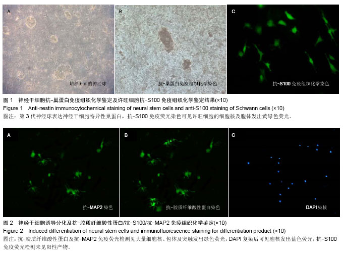
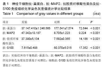
方法:取幼羊脑组织分离神经干细胞后于神经干细胞全培养液中悬浮培养,后期行抗-巢蛋白一抗免疫组织化学染色鉴定,同时取坐骨神经分离培养许旺细胞;以血清诱导干细胞体外诱导分化后行抗-S100一抗、抗-胶质纤维酸性蛋白一抗、抗-MAP2一抗染色分别标记许旺细胞及神经胶质细胞,以未添加一抗做对照,计算图像灰度值与对照组进行统计学比较。

结果与结论:成功分离培养抗-巢蛋白染色阳性神经球及抗-S100染色阳性许旺细胞,后期神经干细胞体外诱导分化得抗-胶质纤维酸性蛋白、抗-MAP2染色阳性细胞,其免疫组织化学染色灰度值显著高于对照组;分化产物抗-S100染色为阴性,其免疫组织化学染色灰度值显著低于对照组。结果表明山羊神经干细胞可经体外诱导分化为神经元及星形胶质细胞,未发现神经干细胞诱导分化为许旺细胞。


中国组织工程研究杂志出版内容重点:干细胞;骨髓干细胞;造血干细胞;脂肪干细胞;肿瘤干细胞;胚胎干细胞;脐带脐血干细胞;干细胞诱导;干细胞分化;组织工程


全文链接:

关键词: 干细胞, 培养, 神经干细胞, 干细胞培养, 体外诱导分化}, 免疫组织化学, 山羊

Abstract:

BACKGROUND: Different types of nerve regulatory factors and glial cells have been reported to exert different roles in the differentiation and maturation of neural stem cells, but culturing neural stem cells in large animals is relatively rarely reported.

OBJECTIVE: To explore the way for culturing goat neural stem cells and to detect the outcome after in vitro differentiation.
METHODS: The neural stem cell was separated and cultured from the newborn goat cerebral cortex and the anti-nestin immunocytochemical staining was performed for cell identification. At the same time, anti-S100 active Schwann cells were gotten from the sciatic nerve. Then in vitro differentiation was preformed and the outcome was detected by the immunocytochemical stain of anti-glial fibrillary acidic protein, anti-microtubule-associated protein 2 and anti-S100. Cells without primary antibodies served as controls. Gray values were calculated and compared.
RESULTS AND CONCLUSION: The Schwann cells were cultured successfully, which were active to the anti-nestin immunocytochemical staining and anti-S100 staining. After differentiation, the products were active to anti-glial fibrillary acidic protein and anti-microtubule-associated protein 2 immunocytochemical stain, but

negative to the anti-S100. And significant difference was found in gray values. The goat neural stem cells and Schwann cells were successfully cultured and identified. After the differentiation, the astrocytes and neurons were detected, but the Schwann cells were not found.


中国组织工程研究杂志出版内容重点:干细胞;骨髓干细胞;造血干细胞;脂肪干细胞;肿瘤干细胞;胚胎干细胞;脐带脐血干细胞;干细胞诱导;干细胞分化;组织工程


全文链接:

Key words: stem cells, neural stem cells, cells, cultured, goats

中图分类号: