| [1] 翁立强,程波.胸腺素促毛囊生长作用研究进展[J]. 国际皮肤性病学杂志, 2007,33(4):244-246.[2] Knop J, App C, Hannappel E. Antibodies in research of thymosin β4: investigation of cross-reactivity and influence of fixatives.Ann N Y Acad Sci. 2012;1270:105-111.[3] Wang X, Yang G, Li S,et al.The Escherichia coli-derived thymosin β4 concatemer promotes cell proliferation and healing wound in mice.Biomed Res Int. 2013;2013:241721.[4] Wei M, Duan D, Liu Y,et al. Increased thymosin β4 levels in the serum and SF of knee osteoarthritis patients correlate with disease severity.Regul Pept. 2013;185:34-36. [5] Xiong Y, Mahmood A, Meng Y,et al.Neuroprotective and neurorestorative effects of thymosin β4 treatment following experimental traumatic brain injury.Ann N Y Acad Sci. 2012; 1270:51-58.[6] Davison G, Brown S.The potential use and abuse of thymosin β-4 in sport and exercise science.J Sports Sci. 2013;31(9): 917-918. [7] Yan B, Singla RD, Abdelli LS,et al. Regulation of PTEN/Akt Pathway Enhances Cardiomyogenesis and Attenuates Adverse Left Ventricular Remodeling following Thymosin β4 Overexpressing Embryonic Stem Cell Transplantation in the Infarcted Heart.PLoS One. 2013;8(9):e75580. [8] Lee SI, Kim DS, Lee HJ,et al.The role of thymosin beta 4 on odontogenic differentiation in human dental pulp cells.PLoS One. 2013;8(4):e61960.[9] Sosne G, Qiu P, Ousler GW 3rd,et al.Thymosin β4: a potential novel dry eye therapy. Ann N Y Acad Sci. 2012;1270: 45-50. [10] Morris DC, Zhang ZG, Zhang J,et al.Treatment of neurological injury with thymosin β4.Ann N Y Acad Sci. 2012;1269: 110-116. [11] Stark C, Taimen P, Tarkia M,et al.Therapeutic potential of thymosin β4 in myocardial infarct and heart failure.Ann N Y Acad Sci. 2012;1269:117-124. [12] Wang WS, Chen PM, Hsiao HL,et al.Overexpression of the thymosin beta-4 gene is associated with malignant progression of SW480 colon cancer cells.Oncogene. 2003; 22(21):3297-3306.[13] Mannherz HG, Hannappel E.The beta-thymosins: intracellular and extracellular activities of a versatile actin binding protein family.Cell Motil Cytoskeleton. 2009;66(10):839-851. [14] Can B, Karagoz F, Yildiz L,et al. Thymosin β4 is a novel potential prognostic marker in gastrointestinal stromal tumors.APMIS. 2012;120(9):689-698. [15] Xiao Y, Chen Y, Wen J,et al.Thymosin β4: a potential molecular target for tumor therapy.Crit Rev Eukaryot Gene Expr. 2012;22(2):109-116.[16] Philp D, Goldstein AL, Kleinman HK.Thymosin beta4 promotes angiogenesis, wound healing, and hair follicle development.Mech Ageing Dev. 2004;125(2):113-115.[17] Philp D, Nguyen M, Scheremeta B,et al.Thymosin beta4 increases hair growth by activation of hair follicle stem cells.FASEB J. 2004;18(2):385-387.[18] Sosne G, Siddiqi A, Kurpakus-Wheater M.Thymosin-beta4 inhibits corneal epithelial cell apoptosis after ethanol exposure in vitro.Invest Ophthalmol Vis Sci. 2004;45(4): 1095-1100.[19] Sribenja S, Wongkham S, Wongkham C,et al. Roles and mechanisms of β-thymosins in cell migration and cancer metastasis: an update.Cancer Invest.2013;31(2):103-110.[20] Fuchs E, Raghavan S.Getting under the skin of epidermal morphogenesis.Nat Rev Genet.2002;3(3):199-209.[21] Huelsken J, Vogel R, Erdmann B,et al.beta-Catenin controls hair follicle morphogenesis and stem cell differentiation in the skin.Cell. 2001;105(4):533-545.[22] Wang LC, Liu ZY, Gambardella L,et al. Regular articles: conditional disruption of hedgehog signaling pathway defines its critical role in hair development and regeneration.J Invest Dermatol. 2000;114(5):901-908.[23] Hwang J, Mehrani T, Millar SE,et al. Dlx3 is a crucial regulator of hair follicle differentiation and cycling.Development. 2008; 135(18):3149-3159. [24] Slominski A, Paus R. Melanogenesis is coupled to murine anagen: toward new concepts for the role of melanocytes and the regulation of melanogenesis in hair growth.J Invest Dermatol. 1993;101(1 Suppl):90S-97S.[25] Alonso L, Fuchs E.The hair cycle.J Cell Sci. 2006;119(Pt 3): 391-393.[26] Hertzog M, van Heijenoort C, Didry D,et al.The beta-thymosin/WH2 domain; structural basis for the switch from inhibition to promotion of actin assembly.Cell. 2004; 117(5):611-623.[27] Huntzicker EG, Oro AE.Controlling hair follicle signaling pathways through polyubiquitination.J Invest Dermatol. 2008; 128(5):1081-1087.[28] Cha HJ, Philp D, Lee SH,et al. Over-expression of thymosin beta 4 promotes abnormal tooth development and stimulation of hair growth.Int J Dev Biol. 2010;54(1):135-140. [29] Jeon BJ, Yang Y, Kyung Shim S, et al. Thymosin beta-4 promotes mesenchymal stem cell proliferation via an interleukin-8-dependent mechanism.Exp Cell Res. 2013; 319(17):2526-2534. [30] Meier N, Langan D, Hilbig H,et al.Thymic peptides differentially modulate human hair follicle growth.J Invest Dermatol. 2012;132(5):1516-1519. [31] Fuchs E, Merrill BJ, Jamora C,et al. At the roots of a never-ending cycle.Dev Cell. 2001;1(1):13-25.[32] DasGupta R, Fuchs E.Multiple roles for activated LEF/TCF transcription complexes during hair follicle development and differentiation.Development. 1999;126(20):4557-4568.[33] 李艳,王冠,于虎.胸腺素β4促进创伤愈合过程中对多种生长因子及新生细胞迁移的作用[J]. 中国组织工程研究与临床康复, 2008,12(11):2143-2147.[34] Shimizu H, Morgan BA.Wnt signaling through the beta-catenin pathway is sufficient to maintain, but not restore, anagen-phase characteristics of dermal papilla cells.J Invest Dermatol. 2004;122(2):239-245.[35] Lowry WE, Blanpain C, Nowak JA,et al. Defining the impact of beta-catenin/Tcf transactivation on epithelial stem cells.Genes Dev. 2005;19(13):1596-1611. [36] Behrens J, von Kries JP, Kühl M,et al. Functional interaction of beta-catenin with the transcription factor LEF-1.Nature. 1996;382(6592):638-642.[37] Roose J, Molenaar M, Peterson J,et al. The Xenopus Wnt effector XTcf-3 interacts with Groucho-related transcriptional repressors.Nature. 1998;395(6702):608-612.[38] Tutter AV, Fryer CJ, Jones KA.Chromatin-specific regulation of LEF-1-beta-catenin transcription activation and inhibition in vitro.Genes Dev. 2001;15(24):3342-3354.[39] Chan SK, Struhl G. Evidence that Armadillo transduces wingless by mediating nuclear export or cytosolic activation of Pangolin.Cell. 2002;111(2):265-280.[40] Zorn AM, Barish GD, Williams BO,et al.Regulation of Wnt signaling by Sox proteins: XSox17 alpha/beta and XSox3 physically interact with beta-catenin.Mol Cell. 1999;4(4): 487-498. |
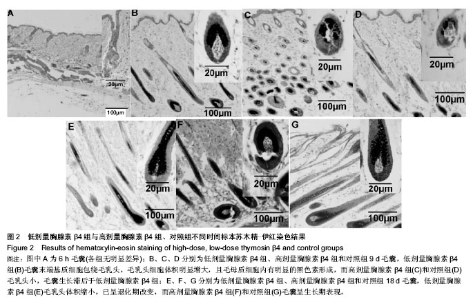
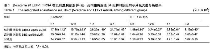
.jpg)