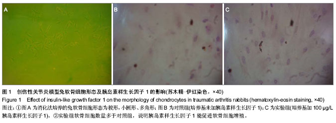
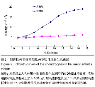
| [1] 陈飞飞,田清业,张祚勇,等.关节软骨缺损修复的研究进展[J].中国骨与关节杂志,2012,1(6):509-512.[2] 朱宝玉,王万春,唐新桥.创伤性关节炎发病机制研究进展[J].国际骨科学杂志,2007,4(28):245-247.[3] 付勤, 肖逸鹏, 王志为,等.生长因子促成年兔关节软骨细胞增殖的研究[J].中国修复重建外科杂志, 2002, 16(4):219- 222.[4] Baek CH, Lee JC, Jung YG,et al.Tissue-engineered cartilage on biodegradable macroporous scaffolds: cell shape and phenotypic expression.Laryngoscope. 2002;112(6): 1050-1055.[5] Baek CH, Ko YJ.Characteristics of tissue-engineered cartilage on macroporous biodegradable PLGA scaffold.Laryngoscope. 2006;116(10):1829-1834.[6] Munirah S, Kim SH, Ruszymah BH,et al.The use of fibrin and poly(lactic-co-glycolic acid) hybrid scaffold for articular cartilage tissue engineering: an in vivo analysis.Eur Cell Mater. 2008;15:41-52.[7] Guerne PA, Blanco F, Kaelin A,et al.Growth factor responsiveness of human articular chondrocytes in aging and development.Arthritis Rheum. 1995;38(7):960-968.[8] Barbero A, Grogan S, Schäfer D,et al.Age related changes in human articular chondrocyte yield, proliferation and post-expansion chondrogenic capacity.Osteoarthritis Cartilage. 2004;12(6):476-484.[9] 方锐,艾力江•阿斯拉,卢勇,等.兔骨性关节炎模型构建及早中晚期的特点[J].中国组织工程研究与临床康复,2010,7(14): 1218-1222.[10] Murray MM, Zurakowski D, Vrahas MS.The death of articular chondrocytes after intra-articular fracture in humans.J Trauma. 2004;56(1):128-131.[11] 田峰,崔学生,张帅,等.改良分步消化法培养原代软骨细胞[J].中国组织工程研究与临床康复,2011,15(20):3633-3635.[12] Lin PM, Chen CT, Torzilli PA.Increased stromelysin-1 (MMP-3), proteoglycan degradation (3B3- and 7D4) and collagen damage in cyclically load-injured articular cartilage.Osteoarthritis Cartilage. 2004;12(6):485-496.[13] Stevens DA, Williams GR.Hormone regulation of chondrocyte differentiation and endochondral bone formation.Mol Cell Endocrinol. 1999;151(1-2):195-204. [14] Mackie EJ, Ahmed YA, Tatarczuch L,et al.Endochondral ossification: how cartilage is converted into bone in the developing skeleton.Int J Biochem Cell Biol. 2008;40(1): 46-62.[15] Chen Y, Ke J, Long X, et al. Insulin-like growth factor-1 boosts the developing process of condylar hyperplasia by stimulating chondrocytes proliferation. Osteoarthritis Cartilage. 2012; 20(4): 279-287. [16] Carlevaro MF, Cermelli S, Cancedda R,et al.Vascular endothelial growth factor (VEGF) in cartilage neovascularization and chondrocyte differentiation: auto-paracrine role during endochondral bone formation.J Cell Sci. 2000;113 ( Pt 1):59-69.[17] Yammani RR, Loeser RF.Extracellular nicotinamide phosphoribosyltransferase (NAMPT/visfatin) inhibits insulin-like growth factor-1 signaling and proteoglycan synthesis in human articular chondrocytes.Arthritis Res Ther. 2012;14(1):R23.[18] Zhang M, Zhou Q, Liang QQ,et al.IGF-1 regulation of type II collagen and MMP-13 expression in rat endplate chondrocytes via distinct signaling pathways.Osteoarthritis Cartilage. 2009;17(1):100-106.[19] 魏立,江莉婷,周琦,等. 大鼠髁突软骨细胞凋亡与胰岛素样生长因子1的影响[J].中国组织工程研究,2013,17(33): 5901-5908.[20] 肖飞,陈聚伍,王福建,等.胰岛素样生长因子-1在家兔骨折愈合过程中的作用[J].中华实验外科杂志,2013,30(9):1936-1938. |

