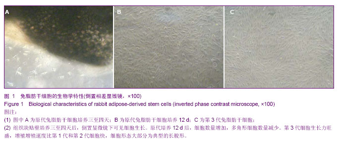
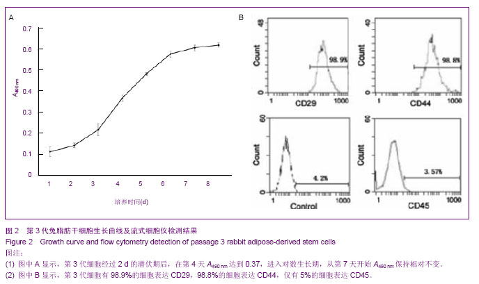
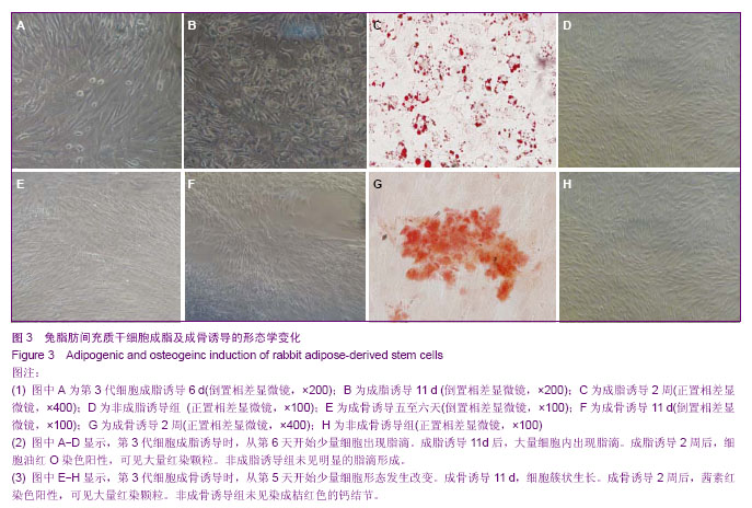
| [1] de Girolamo L, Arrigoni E, Stanco D, et al. Role of autologous rabbit adipose-derived stem cells in the early phases of the repairing process of critical bone defects. J Orthop Res. 2011; 29(1):100-108.[2] Arrigoni E, de Girolamo L, Di Giancamillo A, et al. Adipose-derived stem cells and rabbit bone regeneration: histomorphometric, immunohistochemical and mechanical characterization. J Orthop Sci. 2013;18(2):331-339.[3] Li H, Xu Y, Fu Q, et al. Effects of multiple agents on epithelial differentiation of rabbit adipose-derived stem cells in 3D culture. Tissue Eng Part A. 2012;18(17-18):1760-1770.[4] Arrigoni E, de Girolamo L, Di Giancamillo A, et al. Adipose-derived stem cells and rabbit bone regeneration: histomorphometric, immunohistochemical and mechanical characterization. J Orthop Sci. 2013;18(2):331-339.[5] Baptista LS, do Amaral RJ, Carias RB, et al. An alternative method for the isolation of mesenchymal stromal cells derived from lipoaspirate samples. Cytotherapy. 2009;11(6):706-715. [6] 彭智,陈崎,贾振华,等.组织块培养法扩增人脂肪源性干细胞的生物学特征鉴定[J].中国组织工程研究, 2010, 14(36):6689-6694.[7] Sunay O, Can G, Cakir Z, et al. Autologous rabbit adipose tissue-derived mesenchymal stromal cells for the treatment of bone injuries with distraction osteogenesis. Cytotherapy. 2013;15(6):690-702.[8] Sgodda M, Aurich H, Kleist S, et al. Hepatocyte differentiation of mesenchymal stem cells from rat peritoneal adipose tissue in vitro and in vivo. Exp Cell Res. 2007;313(13):2875-2886.[9] Toghraie F, Razmkhah M, Gholipour MA, et al. Scaffold-free adipose-derived stem cells (ASCs) improve experimentally induced osteoarthritis in rabbits. Arch Iran Med. 2012; 15(8): 495-499.[10] 庞荣清,何洁,李福兵,等.一种简单的人脐带间充质干细胞分离培养方法[J].中华细胞与干细胞杂志:电子版,2011,1(2):162-167.[11] Yang X, Qu L, Wang X, et al. Plasticity of epidermal adult stem cells derived from adult goat ear skin. Mol Reprod Dev. 2007;74(3):386-396.[12] Spath L, Rotilio V, Alessandrini M, et al. Explant-derived human dental pulp stem cells enhance differentiation and proliferation potentials. J Cell Mol Med. 2010;14(6B):1635- 1644.[13] Priya N, Sarcar S, Majumdar AS, et al. Explant culture: a simple, reproducible, efficient and economic technique for isolation of mesenchymal stromal cells from human adipose tissue and lipoaspirate. J Tissue Eng Regen Med. 2012; 1-9.[14] Ishige I, Nagamura-Inoue T, Honda MJ, et al. Comparison of mesenchymal stem cells derived from arterial, venous, and Wharton's jelly explants of human umbilical cord. Int J Hematol. 2009;90(2):261-269.[15] Miranville A, Heeschen C, Sengenès C, et al. Improvement of postnatal neovascularization by human adipose tissue-derived stem cells. Circulation. 2004;110(3):349-355.[16] Yoshimura K, Shigeura T, Matsumoto D, et al. Characterization of freshly isolated and cultured cells derived from the fatty and luid portions of liposuction aspirates. J Cell Physiol. 2006;208(1): 64-76.[17] Brzoska M, Geiger H, Gauer S, et al. Epithelial differentiation of human adipose tissue-derived adult stem cells. Biochem Biophys Res Commun. 2005;330(1):142-150.[18] Sotiropoulou PA, Perez SA, Salagianni M, et al. Characterization of the optimal culture conditions for clinical scale production of human mesenchymal stem cells. Stem Cells. 2006;24(2):462-471.[19] Ou Y, Yuan XD, Cai YN, et al. A novel ethanol-based method to induce differentiation of adipose-derived stromal cel ls into astrocytes. Neural Regen Res. 2011;6(10): 738-743.[20] Baer PC, Griesche N, Luttmann W, et al. Human adipose-derived mesenchymal stem cells in vitro: evaluation of an optimal expansion medium preserving stemness. Cytotherapy. 2010;12(1):96-106.[21] Campagnoli C, Roberts IA, Kumar S, et al. Identification of mesenchymal stem/progenitor cells in human firsttrim ester fetal blood, liver and bone marrow. Blood. 2001;98(8): 2396-2402.[22] Cai YN, Yuan XD, Ou Y, et al. Apoptosis during β-mercaptoethanol-induced differentiation of adult adipose-derived stromal cel ls into neurons. Neural Regen Res. 2011;6(10):750-755.[23] Gronthos S, Franklin DM, Leddy HA, et al. Surface protein characterization of human adipose tissue-derived stromal cells. J Cell Physiol. 2001;189(1):54-63.[24] Gu F, Gao JH, Lu F. Culture of the single cell clones of human adipose-derived stem cells and identification of their surface antigen expressions. Nan Fang Yi Ke Da Xue Xue Bao. 2008; 28(6):1067-1069. [25] Zuk PA, Zhu M, Ashjian P, et al. Human adipose tissue is a source of multipotent stem cells. Mol Biol Cell. 2002;13(12): 4279-4295.[26] Dominici M, Le Blanc K, Mueller I, et al. Minimal criteria for de?ning multipotent mesenchymal stromal cells. The international Society for Cellular Therapy position statement. Cytotherapy. 2006;8(4):315-317.[27] Varma MJ, Breuls RG, Schouten TE, et al. Phenotypical and functional characterization of freshly isolated adipose tissue-derived stem cells. Stem Cells Dev. 2007; 16(1): 91-104.[28] Lindroos B, Suuronen R, Miettinen S. The potential of adipose stem cells in regenerative medicine. Stem Cell Rev. 2011;7(2):269-291.[29] Baer PC, Geiger H. Adipose-derived mesenchymal stromal/stem cells: tissue localization, characterization, and heterogeneity. Stem Cells Int. 2012;2012:812693.[30] Xiao J, Yang X, Jing W, et al. Adipogenic and osteogenic differentiation of Lin(-)CD271(+)Sca-1(+) adipose-derived stem cells. Mol Cell Biochem, 2013. [Epub ahead of print].[31] Zuk P. Adipose-Derived Stem Cells in Tissue Regeneration: A Review. ISRN Stem Cells. 2013;2013:1-35.[32] Yuan XD, Cai YN, Ou Y, et al. Adult adipose-derived stromal cells differentiate into neurons with normal electrophysiological functions. Neural Regen Res. 2011;6(34): 2681-2686.[33] 王清富,陈庄洪,蔡贤华,等.兔脂肪干细胞的多向诱导分化[J].中国组织工程研究, 2012, 16(49):9162-9167.[34] Cawthorn WP, Scheller EL, MacDougald OA. Adipose tissue stem cells: the great WAT hope.Trends Endocrinol Metab. 2012;23(6):270-277.[35] Macotela Y, Emanuelli B, Mori MA, et al. Intrinsic differences in adipocyte precursor cells from different white fat depots. Diabetes. 2012;61(7):1691-1699.[36] Suga H, Matsumoto D, Eto H, et al. Functional implications of CD34 expression in human adipose-derived stem/progenitor cells. Stem Cells Dev. 2009;18(8):1201-1209.[37] Rowlands AS, George PA, Cooper-White JJ. Directing osteogenic and myogenic differentiation of MSCs: interplay of stiffness and adhesive ligand presentation.Am J Physiol Cell Physiol. 2008;295(4):C1037-1044.[38] Buehrer BM, Cheatham B. Isolation and characterization of human adipose-derived stem cells for use in tissue engineering. Methods Mol Biol. 2013;1001:1-11. |
.jpg)