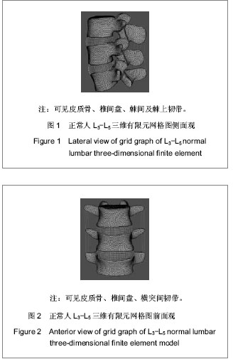
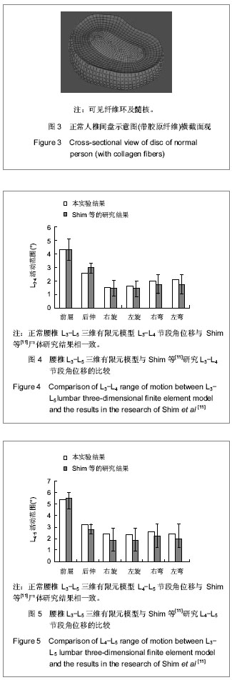
| [1] Belytschko T, Kulak RF, Schultz AB,et al.Finite element stress analysis of an intervertebral disc. J Biomech. 1974;7(3): 277-285.[2] Chen SH, Lin SC, Tsai WC,et al. Biomechanical comparison of unilateral and bilateral pedicle screws fixation for transforaminal lumbar interbody fusion after decompressive surgery -- a finite element analysis.BMC Musculoskelet Disord. 2012;13:72.[3] Lo CC, Tsai KJ, Chen SH,et al. Biomechanical effect after Coflex and Coflex rivet implantation for segmental instability at surgical and adjacent segments: a finite element analysis.Comput Methods Biomech Biomed Engin. 2011;14(11):969-978.[4] Lee KK, Teo EC, Fuss FK,et al. Finite-element analysis for lumbar interbody fusion under axial loading.IEEE Trans Biomed Eng. 2004;51(3):393-400.[5] Schmidt H, Heuer F, Simon U,et al.Application of a new calibration method for a three-dimensional finite element model of a human lumbar annulus fibrosus.Clin Biomech (Bristol, Avon). 2006;21(4):337-344.[6] Shirazi-Adl A, Ahmed AM, Shrivastava SC. Mechanical response of a lumbar motion segment in axial torque alone and combined with compression.Spine (Phila Pa 1976). 1986;11(9):914-927.[7] White Ⅲ AA, Panjabi MM. Clinical Biomechanics of the Spine. Philadelphia: J.B. Lippincott, 1990.[8] Polikeit A, Ferguson SJ, Nolte LP,et al.Factors influencing stresses in the lumbar spine after the insertion of intervertebral cages: finite element analysis.Eur Spine J. 2003;12(4):413-420.[9] Goto K, Tajima N, Chosa E,et al.Effects of lumbar spinal fusion on the other lumbar intervertebral levels (three- dimensional finite element analysis).J Orthop Sci. 2003; 8(4):577-584.[10] Goel VK, Monroe BT, Gilbertson LG,et al.Interlaminar shear stresses and laminae separation in a disc. Finite element analysis of the L3-L4 motion segment subjected to axial compressive loads.Spine (Phila Pa 1976). 1995;20(6):689-698.[11] Shim CS, Park SW, Lee SH,et al. Biomechanical evaluation of an interspinous stabilizing device, Locker.Spine (Phila Pa 1976). 2008;33(22):E820-E827.[12] Goel VK, Gilbertson LG. Applications of the finite element method to thoracolumbar spinal research--past, present, and future.Spine (Phila Pa 1976). 1995;20(15):1719-1727.[13] Yoganandan N, Kumaresan SC, Voo L,et al. Finite element modeling of the C4-C6 cervical spine unit.Med Eng Phys. 1996;18(7):569-574.[14] Tang S, Meng X.Does disc space height of fused segment affect adjacent degeneration in ALIF? A finite element study.Turk Neurosurg. 2011;21(3):296-303.[15] 刘勇,田青,李国庆,等.骨质疏松腰椎三维有限元模型的建立[J].中华实验外科杂志,2013,30(1):161-162.[16] 黄菊英,李海云,菅凤增,等.腰椎间盘突出症有限元模型的建立与分析[J].中国现代神经疾病杂志,2012,12(4):394-398. [17] 朱立新,王健,曹延林,等.基于CT数据构建人体腰骶椎的有限元模型[J].中国组织工程研究,2012,6(30):5511-5515.[18] 付裕,阮狄克,霍洪军,等.基于CT扫描及CAD技术建立下腰椎三维有限元模型[J].中国组织工程研究与临床康复,2011,15(9): 1557-1561.[19] Yoganandan N, Myklebust JB, Ray G,et al. Mathematical and finite element analysis of spine injuries.Crit Rev Biomed Eng. 1987;15(1):29-93.[20] Yoganandan N, Kumaresan S, Voo L,et al.Finite element model of the human lower cervical spine: parametric analysis of the C4-C6 unit.J Biomech Eng. 1997 ;119(1):87-92.[21] Yoganandan N, Kumaresan S, Voo L,et al.Finite element applications in human cervical spine modeling.Spine (Phila Pa 1976). 1996;21(15):1824-1834.[22] Yoganandan N, Kumaresan S, Pintar FA. Biomechanics of the cervical spine Part 2. Cervical spine soft tissue responses and biomechanical modeling.Clin Biomech (Bristol, Avon). 2001; 16(1):1-27.[23] Schultz AB, Warwick DN, Berkson MH, et al. Mechanical Properties of Human Lumbar Spine Motion Segments-Part I: Responses in Flexion, Extension, Lateral Bending, and Torsion. J Biomech Eng. 1979; 101:46-52. [24] Panjabi MM, Krag MH, Chung TQ. Effects of disc injury on mechanical behavior of the human spine.Spine (Phila Pa 1976). 1984;9(7):707-713.[25] Tencer AF, Ahmed AM, Burke DL.Some static mechanical properties of the lumbar intervertebral joint, intact and injured.J Biomech Eng. 1982;104(3):193-201.[26] Shirazi-Adl A, Ahmed AM, Shrivastava SC. A finite element study of a lumbar motion segment subjected to pure sagittal plane moments.J Biomech. 1986;19(4):331-350.[27] Goel VK, Kim YE, Lim TH,et al. An analytical investigation of the mechanics of spinal instrumentation.Spine (Phila Pa 1976). 1988;13(9):1003-1011.[28] Vadapalli S, Sairyo K, Goel VK,et al.Biomechanical rationale for using polyetheretherketone (PEEK) spacers for lumbar interbody fusion-A finite element study.Spine (Phila Pa 1976). 2006;31(26):E992-998.[29] Tsuang YH, Chiang YF, Hung CY,et al. Comparison of cage application modality in posterior lumbar interbody fusion with posterior instrumentation--a finite element study.Med Eng Phys. 2009;31(5):565-570.[30] Zhong ZC, Wei SH, Wang JP,et al.Finite element analysis of the lumbar spine with a new cage using a topology optimization method.Med Eng Phys. 2006;28(1):90-98.[31] Chen SH, Lin SC, Tsai WC,et al. Biomechanical comparison of unilateral and bilateral pedicle screws fixation for transforaminal lumbar interbody fusion after decompressive surgery -- a finite element analysis.BMC Musculoskelet Disord. 2012;13:72.[32] Adam C, Pearcy M, McCombe P.Stress analysis of interbody fusion--finite element modelling of intervertebral implant and vertebral body.Clin Biomech (Bristol, Avon). 2003;18(4): 265-272.[33] Xu H, Tang H, Guan X,et al.Biomechanical comparison of posterior lumbar interbody fusion and transforaminal lumbar interbody fusion by finite element analysis.Neurosurgery. 2013;72(1 Suppl Operative):21-26.[34] 马金梁,李孝林,邓忠良.一种新型椎间融合器用于腰椎融合的有限元模型建立[J].上海医学,2012,35(11):960-963.[35] 马辉,赵杰,侯铁胜,等.腰椎滑脱后路融合术式有限元模型的建立[J].脊柱外科杂志,2008,6(3):132-136. |
.jpg)