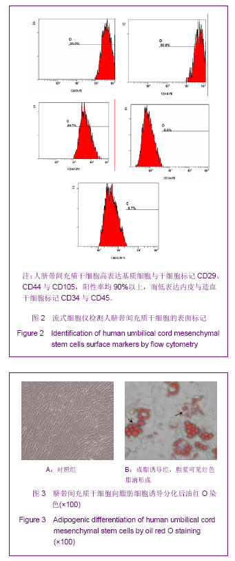
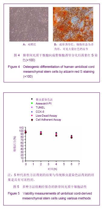
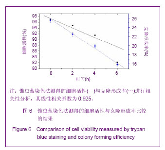
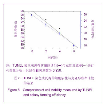
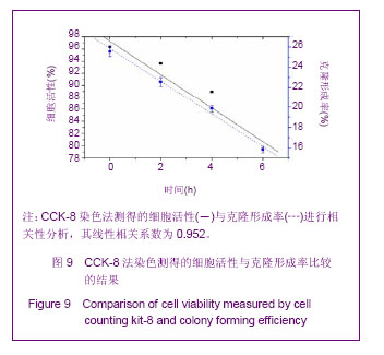
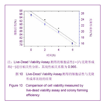
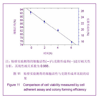
| [1] Lee JW, Fang X, Krasnodembskaya A, et al. Concise review: Mesenchymal stem cells for acute lung injury: role of paracrine soluble factors. Stem Cells. 2011;29(6):913-919.[2] Semenov OV, Koestenbauer S, Riegel M, et al. Multipotent mesenchymal stem cells from human placenta: critical parameters for isolation and maintenance of stemness after isolation. Am J Obstet Gynecol. 2010;202(2):193.e1- 193.e13.[3] Tran CT, Huynh DT, Gargiulo C,et al. In vitro culture of Keratinocytes from human umbilical cord blood mesenchymal stem cells: the Saigonese culture. Cell Tissue Bank. 2011; 12(2):125-133.[4] Can A, Balci D. Isolation, culture, and characterization of human umbilical cord stroma-derived mesenchymal stem cells. Methods Mol Biol. 2011;698:51-62.[5] Singleton PA, Salgia R, Moreno-Vinasco L,et al. CD44 regulates hepatocyte growth factor-mediated vascular integrity. Role of c-Met, Tiam1/Rac1, dynamin 2, and cortactin. J Biol Chem. 2007;282(42):30643-30657.[6] Mei SH, McCarter SD, Deng Y, et al. Prevention of LPS-induced acute lung injury in mice by mesenchymal stem cells overexpressing angiopoietin 1. PLoS Med. 2007;4(9):e269.[7] Manning E, Pham S, Li S, et al. Interleukin-10 delivery via mesenchymal stem cells: a novel gene therapy approach to prevent lung ischemia-reperfusion injury. Hum Gene Ther. 2010;21(6):713-727.[8] Olson AL, Swigris JJ. Idiopathic pulmonary fibrosis: diagnosis and epidemiology. Clin Chest Med. 2012;33(1):41-50.[9] Cho PS, Messina DJ, Hirsh EL,et al. Immunogenicity of umbilical cord tissue derived cells. Blood. 2008;111(1):430- 438.[10] La Rocca G, Anzalone R, Corrao S, et al. Isolation and characterization of Oct-4+/HLA-G+ mesenchymal stem cells from human umbilical cord matrix: differentiation potential and detection of new markers. Histochem Cell Biol. 2009;131(2): 267-282.[11] Venkataramana NK, Kumar SK, Balaraju S,et al. Open-labeled study of unilateral autologous bone-marrow-derived mesenchymal stem cell transplantation in Parkinson's disease.Transl Res. 2010;155(2):62-70.[12] Garzón I, Pérez-Köhler B, Garrido-Gómez J, et al. Evaluation of the cell viability of human Wharton's jelly stem cells for use in cell therapy.Tissue Eng Part C Methods. 2012;18(6): 408-419.[13] Rodriguez-Morata A, Garzon I, Alaminos M, et al. Cell viability and prostacyclin release in cultured human umbilical vein endothelial cells. Ann Vasc Surg. 2008;22(3):440-448.[14] Mazaheri Z, Movahedin M, Rahbarizadeh F, et al. Different doses of bone morphogenetic protein 4 promote the expression of early germ cell-specific gene in bone marrow mesenchymal stem cells. In Vitro Cell Dev Biol Anim. 2011; 47(8):521-525. [15] Sadan O, Shemesh N, Barzilay R,et al. Migration of neurotrophic factors-secreting mesenchymal stem cells toward a quinolinic acid lesion as viewed by magnetic resonance imaging.Stem Cells. 2008;26(10):2542-2551.[16] Seeger FH, Tonn T, Krzossok N, et al. Cell isolation procedures matter: a comparison of different isolation protocols of bone marrow mononuclear cells used for cell therapy in patients with acute myocardial infarction. Eur Heart J. 2007;28(6):766-772.[17] Mihu CM, Mihu D, Costin N, et al. Isolation and characterization of stem cells from the placenta and the umbilical cord. Rom J Morphol Embryol. 2008;49(4):441-446.[18] Horwitz EM, Le Blanc K, Dominici M, et al. Clarification of the nomenclature for MSC: The International Society for Cellular Therapy position statement. Cytotherapy. 2005;7(5):393-395.[19] Heng BC, Cowan CM, Basu S.Temperature and calcium ions affect aggregation of mesenchymal stem cells in phosphate buffered saline.Cytotechnology. 2008;58(2):69-75.[20] 卢润章,赵经慧,程梅,等.人脐带间充质干细胞体外培养的生物学特性研究[J].黑龙江医学,2013,37(2):81-83.[21] 张芬熙,洪艳,梁文妹.人脐带间充质干细胞的分离培养及超微结构特点研究[J].贵阳医学院学报,2013,38(1):5-9,15.[22] 赵庆华,祝加学,王雷,等.人脐带间充质干细胞的生物学特性及向软骨细胞、骨细胞分化实验研究[J].中华医学杂志,2011,91(5): 317-321.[23] 孙丽,于丽,张华芳,等.人脐带间充质干细胞的分离培养及生物学特性[J].解剖科学进展,2011,17(2):131-134.[24] Mazaheri Z, Movahedin M, Rahbarizadeh F,et al. Different doses of bone morphogenetic protein 4 promote the expression of early germ cell-specific gene in bone marrow mesenchymal stem cells.In Vitro Cell Dev Biol Anim. 2011; 47(8):521-525.[25] Guo RM, Cao N, Zhang F,et al.Controllable labelling of stem cells with a novel superparamagnetic iron oxide-loaded cationic nanovesicle for MR imaging. Eur Radiol. 2012;22(11): 2328-2337.[26] Sun JH, Zhang YL, Qian SP,et al.Assessment of biological characteristics of mesenchymal stem cells labeled with superparamagnetic iron oxide particles in vitro.Mol Med Rep. 2012;5(2):317-320.[27] Mascotti K, McCullough J, Burger SR. HPC viability measurement: trypan blue versus acridine orange and propidium iodide.Transfusion. 2000;40(6):693-696.[28] 刘阳,赖翼,李敏惠,等.流式细胞术检测细胞存活率的方法学建立[J].国际检验医学杂志,2011,32(15):1663-1664.[29] Chan LL, Wilkinson AR, Paradis BD, et al. Rapid image-based cytometry for comparison of fluorescent viability staining methods. J Fluoresc. 2012;22(5):1301-1311.[30] van der Bogt KE, Schrepfer S, Yu J, et al. Comparison of transplantation of adipose tissue- and bone marrow-derived mesenchymal stem cells in the infarcted heart. Transplantation. 2009;87(5):642-652.[31] Assmus B, Tonn T, Seeger FH, et al. Red blood cell contamination of the final cell product impairs the efficacy of autologous bone marrow mononuclear cell therapy. J Am Coll Cardiol. 2010;55(13):1385-1394.[32] Song H, Cha MJ, Song BW, et al. Reactive oxygen species inhibit adhesion of mesenchymal stem cells implanted into ischemic myocardium via interference of focal adhesion complex. Stem Cells. 2010;28(3):555-563.[33] Sarugaser R, Lickorish D, Baksh D,et al. Human umbilical cord perivascular (HUCPV) cells: a source of mesenchymal progenitors.Stem Cells. 2005;23(2):220-229. |


.jpg)