中国组织工程研究 ›› 2013, Vol. 17 ›› Issue (32): 5772-5777.doi: 10.3969/j.issn.2095-4344.2013.32.005

• 脐带脐血干细胞 umbilical cord blood stem cells • 上一篇    下一篇

人脐带间充质干细胞与肝细胞共培养可分化为肝样细胞

李  华1,文  峰2,漆仲春1,周进军1,朱亚杰1,程  朋1,魏  东1,苏晓妹1,谭  勇1,彭晶晶1,罗巧丽1,李  东1,张  涛1   

  1. 1解放军成都军区总医院肿瘤诊治中心,四川省成都市  610083
    2成都医学院第一附属医院肿瘤科,四川省成都市   610513
  • 收稿日期:2012-09-11 修回日期:2012-10-17 出版日期:2013-08-06 发布日期:2013-08-06
  • 通讯作者: 张涛,硕士,主任医师,解放军成都军区总医院肿瘤诊治中心 ,四川省成都市 610083 zhangtao269@126.com
  • 作者简介:李华☆,女,1971年生,四川省成都市人,汉族,2009年解放军第三军医大学毕业,博士,副主任技师,主要从事肿瘤免疫的研究。 Huali99@gmail.com
  • 基金资助:

    四川省科技厅面上项目(2009JY0016,2012JY0031);四川省科技支撑计划(2012SZ0058)。

Human umbilical cord-derived mesenchymal stem cells co-cultured with hepatocytes can differentiate into hepatocyte-like cells

Li Hua1, Wen Feng2, Qi Zhong-chun1, Zhou Jin-jun1, Zhu Ya-jie1, Cheng Peng1, Wei Dong1, Su Xiao-mei1, Tan Yong1, Peng Jing-jing1, Luo Qiao-li1, Li Dong1, Zhang Tao1   

  1. 1Department of Oncology, General Hospital of Chengdu Military Region of PLA, Chengdu  610083, Sichuan Province, China
    2First Affiliated Hospital of Chengdu Medical College, Chengdu  610513, Sichuan Province, China
  • Received:2012-09-11 Revised:2012-10-17 Online:2013-08-06 Published:2013-08-06
  • Contact: Zhang Tao, Master, Chief physician, Department of Oncology, General Hospital of Chengdu Military Region of PLA, Chengdu 610083, Sichuan Province, China zhangtao269@126.com
  • About author:Li Hua☆, M.D., Associate chief technician, Department of Oncology, General Hospital of Chengdu Military Region of PLA, Chengdu 610083, Sichuan Province, China Huali99@gmail.com
  • Supported by:

    General Project of Sichuan Science and Technology Deparment, No.2009JY0016*, 2012JY0031*; Key Technology Project of Sichuan Province, No. 2012SZ0058*

摘要:

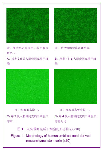
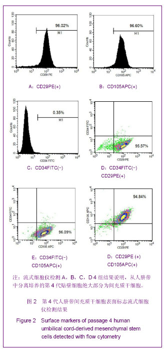
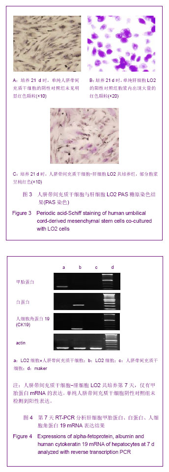
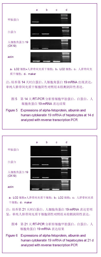
背景:文献报道,从骨髓与脐带中分离获得的间充质干细胞可在体外连续传代培养,仍保持干细胞的特性,并在多种细胞因子的“鸡尾酒式”诱导下分化为肝细胞样细胞。
目的:进一步验证人脐带间充质干细胞在体外正常人肝细胞共培养体系下是否可分化为肝细胞并探讨其分化方法。
方法:采用贴壁法,从脐带中分离培养间充质干细胞,流式细胞仪检测脐带间充质干细胞表面标志。人肝细胞LO2细胞与人脐带间充质干细胞建立共培养体系,不添加外源诱导因子,分别于第7,14,21天,通过RT-PCR 法检测肝细胞特异标志物甲胎蛋白、白蛋白、人细胞角蛋白19 mRNA的表达,糖原染色进行功能鉴定。
结果与结论:从人脐带中可分离得到贴壁生长的间充质干细胞,其中CD29+细胞比例为96.02%,CD105+细胞比例为96.6%,CD34-细胞比例为99.65%,CD105+CD29+双阳性细胞比例为94.84%。与LO2细胞共培养后第7天仅有甲胎蛋白阳性表达;第14天表达白蛋白、人细胞角蛋白19,第21天时,LO2与人脐带间充质干细胞共培养组未出现甲胎蛋白表达;人细胞角蛋白19和白蛋白的表达比第14天略有增强。共培养21 d后,糖原染色呈阳性。结果证实,无需额外添加外源诱导因子,脐带间充质干细胞可在人正常肝细胞共培养的微环境中,向正常肝细胞分化。

关键词: 干细胞, 脐带脐血干细胞, 脐带间充质干细胞, 肝细胞, 共培养, 细胞分化, 细胞培养, 肝样细胞, 甲胎蛋白, 白蛋白, 人细胞角蛋白19, 省级基金, 干细胞图片文章

Abstract:

BACKGROUND: The studies have shown that the mesenchymal stem cells derived from bone marrow and umbilical cord can be continuously cultured in vitro, and maintain the characteristics of stem cells. The mesenchymal stem cells can differentiate into hepatocyte-like cells after “cocktail” induction by various cytokines.
OBJECTIVE: To further identify whether umbilical cord-derived mesenchymal stem cells in vitro co-cultured with normal hepatocytes can differentiate into hepatocyte-like cells, and to investigate the differentiation method.
METHODS: Mesenchymal stem cells were isolated from human umbilical cord with adherent method, and the surface markers of umbilical cord-derived mesenchymal stem cells were detected with flow cytometry. The umbilical cord-derived mesenchymal stem cells were co-cultured with liver LO2 cells without adding exogenous inducers. The expressions of alpha-fetoprotein, albumin and human cytokeratin 19 mRNA of hepatocyte specific markers were detected with reverse transcription PCR at 7, 14 and 21 days after culture, and periodic acid-Schiff staining was used to identify the functions.
RESULTS AND CONCLUSION: Mesenchymal stem cells could isolated from human umbilical cord successfully, showing fibroblastic morphology and adherent cell characterization. Among these cells, 96.02% cells were CD29 positive cells and 96.6% cells were CD105 positive cells. The percentage of CD34 negative cells was 99.65%. The percentage of CD105+CD29+ double positive cells was 94.84%. The mRNA of alpha-fetoprotein was found on the 7th day after co-cultured with LO2 cells, and the mRNA of albumin and human cytokeratin 19 were found on the 14th day. After co-cultured for 21 days, the alpha-fetoprotein mRNA could not be observed in the co-culture group. The expressions of albumin and human cytokeratin 19 were increased at 14 days. After co-cultured for 21 days, the glycogen staining was positive. Umbilical cord-derived mesenchymal stem cells can differentiate into hepatocyte-like cells after co-cultured with normal hepatocytes.

Key words: stem cells, umbilical cord/umbilical blood stem cells, umbilical cord-derived mesenchymal stem cells, hepatocytes, co-culture, cell differentiation, cell culture, hepatocyte-like cells, alpha-fetoprotein, albumin, human cytokeratin 19, provincial grants-supported paper, stem cell photographs-containing paper

中图分类号: