中国组织工程研究 ›› 2013, Vol. 17 ›› Issue (29): 5290-5295.doi: 10.3969/j.issn.2095-4344.2013.29.005

• 组织工程口腔材料 tissue-engineered oral materials • 上一篇    下一篇

小型猪牙周膜干细胞在羟基磷灰石支架材料上的生长

马路平1,钟良军2,张源明3,张 远4,张鹏涛4   

  1. 新疆医科大学第一附属医院,1口腔科,3心脏中心,新疆维吾尔自治区乌鲁木齐市  830054;2杭州师范大学临床医学院口腔医学系,浙江省杭州市  310015;4杭州师范大学附属医院口腔科,浙江省杭州市 310015
  • 收稿日期:2013-04-16 修回日期:2013-06-07 出版日期:2013-07-22 发布日期:2013-07-22
  • 通讯作者: 张源明,教授,博士生导师,新疆医科大学第一附属医院心脏中心,新疆维吾尔自治区乌鲁木齐市 830054 zymdxx@163.com
  • 作者简介:马路平★,女,1982年生,汉族,湖南省人,新疆医科大学第一附属医院在读硕士,主要从事牙周病的防治工作。 yayamlp@qq.com
  • 基金资助:

    自治区科技支疆项目(201091144)

Periodontal ligament cells from miniature swine grow on a hydroxyapatite scaffold

Ma Lu-ping1, Zhong Liang-jun2, Zhang Yuan-ming3, Zhang Yuan4, Zhang Peng-tao4   

  1. 1Department of Stomatology, First Affiliated Hospital of Xinjiang Medical University, Urumqi  830054, Xinjiang Uygur Autonomous Region, China; 2Department of Stomatology, Clinical College of Hangzhou Normal University, Hangzhou  310015, Zhejiang Province, China; 3Department of Cardiology, First Affiliated Hospital of Xinjiang Medical University, Urumqi  830054, Xinjiang Uygur Autonomous Region, China; 4Department of Stomatology, Affiliated Hospital of Hangzhou Normal University, Hangzhou  310015, Zhejiang Province, China
  • Received:2013-04-16 Revised:2013-06-07 Online:2013-07-22 Published:2013-07-22
  • Contact: Zhang Yuan-ming, Professor, Doctoral supervisor, Department of Cardiology, First Affiliated Hospital of Xinjiang Medical University, Urumqi 830054, Xinjiang Uygur Autonomous Region, China zymdxx@163.com
  • About author:Ma Lu-ping★, Studying for master’s degree, Department of Stomatology, First Affiliated Hospital of Xinjiang Medical University, Urumqi 830054, Xinjiang Uygur Autonomous Region, China yayamlp@qq.com
  • Supported by:

    Project of Science and Technology Supporting to Xinjiang Uygur Autonomous Region, No. 201091144*

摘要:

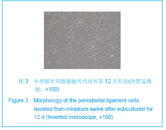
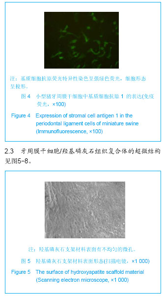
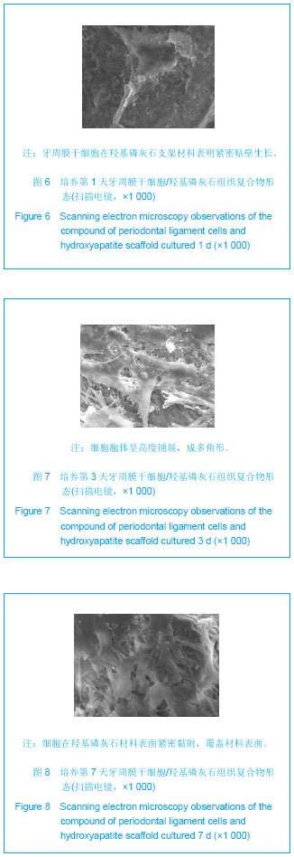
背景:牙周组织工程技术为修复牙周炎骨组织缺损提供新思路和新方法。
目的:体外培养小型猪牙周膜干细胞,并与羟基磷灰石进行骨组织构建,研究其与羟基磷灰石支架材料的生物相容性。
方法:采用组织块法获得小型猪牙周膜干细胞,用免疫荧光法检测细胞中基质细胞抗原1的表达。取第3代小型猪牙周膜干细胞接种在羟基磷灰石支架材料上复合培养,分别在培养第1,3,7天用扫描电镜观察细胞在羟基磷灰石上的生长情况。
结果与结论:原代培养的小型猪牙周膜干细胞生长良好,细胞中基质细胞抗原1免疫荧光呈阳性。扫描电镜结果表明,培养第1,3,7天,牙周膜干细胞在羟基磷灰石上生长良好。证实,实验采用组织块法成功体外分离培养了小型猪牙周膜干细胞,可在羟基磷灰石支架材料上良好的生长。

关键词: 生物材料, 组织工程口腔材料, 牙周膜干细胞, 体外培养, 羟基磷灰石, 扫描电镜, 小型猪, 省级基金

Abstract:

BACKGROUND: Periodontal tissue engineering technology provides new ideas and new ways for periodontitis-induced bone defect repair.
OBJECTIVE: To develop a culture model for the periodontal ligament cells of miniature swine, which was constructed with hydroxyapatite, to investigate the biocompatibility with hydroxyapatite.
METHODS: Periodontal ligament cells from miniature swine were harvested by using tissue explant method. Immunofluorescence was used to detect the expression of stromal cell antigen 1 in the periodontal ligament cells of miniature swine. The third passage cells were co-cultured with a three-dimension hydroxyapatite scaffold, and the biological characteristics of the cells were observed under a scanning electron microscope at days 1, 3, 7 of co-culture.
RESULTS AND CONCLUSION: The pirmary miniature swine periodontal ligament cells grew well, and they were positive for stromal cell antigen 1. Under the scanning electron microscope, the periodontal ligament cells of miniature swine grew well on the hydroxyapatite scaffold at days 1, 3, 7 of co-culture. These prove that the miniature swine periodontal ligament cells, which can be separated using tissue explant method and cultured successfully in vitro, can grow well on the hydroxyapatite scaffold.

Key words: biomaterials, tissue-engineered oral materials, periodontal ligament cells, in vitro culture, hydroxyapatite, scanning electron microscope, miniature swine, provincial grants-supported paper

中图分类号: