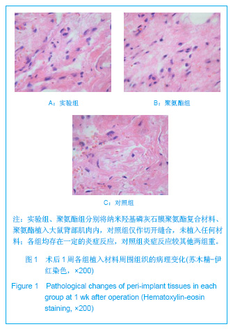
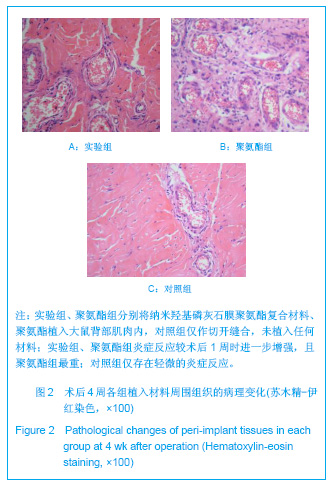
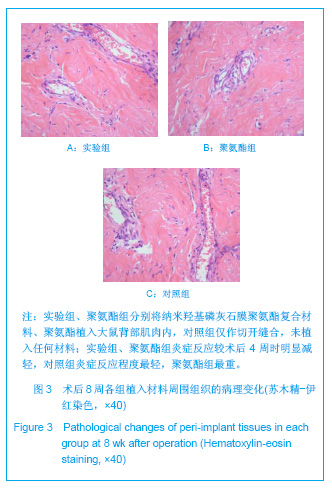
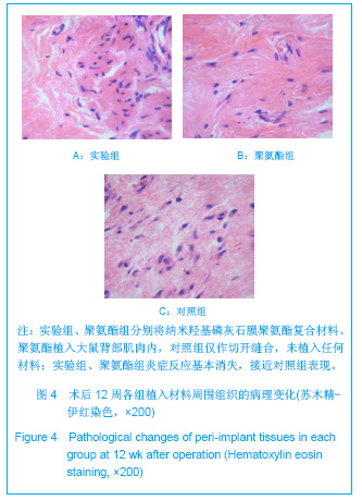
| [1] Devesa JM,Rey A,Hervas PL,et al. Artificial anal sphincter: complications and functional results of a large personal series. Dis Colon Rectum.2002;45(9):1154-1163.[2] Ruff CA, Vujevich JJ,Goldberg LH. Polyurethane dressing assisted epidermal suturing minimizes postoperative wound care.J Drugs Dermatol.2008; 7(7):675-677.[3] Tang JL,Xi TW.Zhongguo Zuzhi Gongcheng Yanjiu yu Linchuang Kangfu. 2007;11(5):936-939. 汤京龙,奚廷斐.纳米羟基磷灰石生物安全性的研究现状[J].中国组织工程研究与临床康复,2007,11(5):936-939.[4] Gui JJ,Chen L,Chen S.hecheng xiangjiao gongye. 2005;28(2): 151-154. 眭建军,陈莉,陈苏.功能聚氨酯材料在生物医学工程中的研究进展及应用[J].合成橡胶工业,2005,28(2):151-154.[5] Guo XB,Huang ZH,Zhang HB,et al.Guangdong Yixue. 2010; 31(1):52-54. 郭雄波,黄宗海,张宏斌,等.纳米羟基磷灰石膜复合材料与小鼠成纤维细胞的相容性[J].广东医学,2010,31(1):52-54.[6] Cheng GC,Yan ZY,Luo L,et al.Anhui Yike Daxue Xuebao. 2007; 42(6):626-629. 程光存,严中亚,罗乐,等. 羟基磷灰石膜复合材料与人血管内皮细胞的相容性[J].安徽医科大学学报,2007,42(6):626-629. [7] Lehur PA,Michot F,Denis P,et al. Results of artificial sphincter in severe anal incontinence. Report of 14 consecutive implantations. Dis Colon Rectum. 1996;39(12):1352-1355.[8] Wu WL,Li J.Zhongguo Zuzhi Gongcheng Yanjiu yu Linchuang Kangfu. 2006;10(45):217-219. 武卫莉,李佳. 硅橡胶与聚氨酯医用材料的生物学特性[J].中国组织工程研究与临床康复,2006,10(45):217-219.[9] Rueda L,Garcia I,Palomares T,et al.The role of reactive silicates on the structure/property relationships and cell response evaluation in polyurethane nanocomposites. J Biomed Mater Res A.2011;97(4):480-489.[10] Chen Y,Webster TJ. Increased osteoblast functions in the presence of BMP-7 short peptides for nanostructured biomaterial applications. J Biomed Mater Res A.2009; 91(1): 296-304.[11] Zhou WY,Wang M,Cheung WL,et al.Synthesis of carbonated hydroxyapatite nanospheres through nanoemulsion.J Mater Sci Mater Med.2008;19(1):103-110.[12] Geary C,Birkinshaw C,Jones E. Characterisation of Bionate polycarbonate polyurethanes for orthopaedic applications.J Mater Sci Mater Med.2008; 19(11):3355-3363.[13] Caracciolo PC,Buffa F,Abraham GA.Effect of the hard segment chemistry and structure on the thermal and mechanical properties of novel biomedical segmented poly (esterurethanes). J Mater Sci Mater Med.2009;20(1):145-155.[14] Meskinfam M,Sadjadi MA,Jazdarreh H,et al. Biocompatibility evaluation of nano hydroxyapatite-starch biocomposites.J Biomed Nanotechnol.2011; 7(3):455-459.[15] Li X,Nan K,Shi S,et al.Preparation and characterization of nano-hydroxyapatite/chitosan cross-linking composite membrane intended for tissue engineering.Int J Biol Macromol. 2011;13(9):423-424.[16] Khan I,Smith N,Jones E,et al.Analysis and evaluation of a biomedical polycarbonate urethane tested in an in vitro study and an ovine arthroplasty model. Part II: in vivo investigation. Biomaterials.2005;26(6):633-643. |