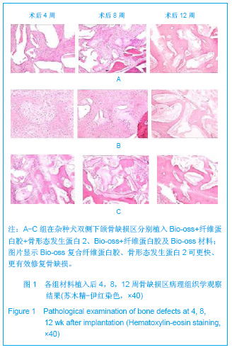
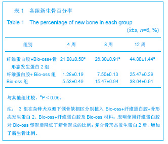
| [1] Marras H.Fibrin seal: the state of the art. J Oral Maxillofac Surg.1985;43(8):605.[2] Wang PY,Bai XF,Wang BW,et al.Zhongguo Chuangxin Yixue. 2011;8(6):9-11. 王平瑜,白雪峰,王保卫,等.人纤维蛋白胶对人肝损伤的止血护创作用[J].中国创新医学,2011,8(6):9-11.[3] Deng QJ,Li YQ,Xie LQ,et al.Zhongguo Shiyong Zhenduan yu Zhiliao Zazhi. 2010;24(12):1212-1213. 邓全军,李永芹,谢立群,等.内镜下金属钛夹联合纤维蛋白胶治疗Dieulafoy病上消化道出血疗效观察[J].中华实用诊断与治疗杂志, 2010,24(12):1212-1213.[4] Huang HJ,Zheng J,Si YG.Zhongguo Yishi Jinxiu Zazhi. 2011; 34(20):71-72. 黄海江,郑军,斯友光. 纤维蛋白胶结合带蒂空肠袢覆盖在十二指肠破裂修补中的应用[J].中国医师进修杂志,2011,34(20): 71-72.[5] Hu XN,Guo N,Zhang DL,et al.Zhonghua Shiyan Waike Zazhi. 2010;27(12):1910-1912. 胡学宁,郭宁,张冬蕾,等.两种组织黏合剂封堵肺创面漏气的研究[J].中华实验外科杂志,2010,27(12):1910-1912.[6] Jin BS,Zheng JW,Jiao Y,et al.Zhongguo Yishi Zazhi. 2008; 10(9):1275-1276. 晋炳申,郑建伟,矫艳,等.纤维蛋白胶预防室间隔缺损术后残余漏发生的研究[J].中国医师杂志,2008,10(9):1275-1276.[7] Matsumoto K,Kohmura E,Kato A,et al.Restoration of small bone defects at craniotomy using autologous bone dust and fibrin glue.Surg Neurol.1998;50(4):344-346.[8] Zhang L,Ding Y,Shao JL.Huaxi Kouqiang Yixue Zazhi. 2011; 29(2):125-128. 张亮,丁寅,邵金陵.骨髓间充质干细胞联合纤维蛋白胶修复大鼠牙槽骨缺损的研究[J].华西口腔医学杂志,2011,29(2):125-128.[9] Cai ZG, Ri G. Zhongguo Zuzhi Gongcheng Yanjiu yu Linchuang Kangfu. 2011;15(17):2933-2936. 蔡志剐,芮钢.纤维蛋白胶复合自体骨髓与人工骨促进脊柱融合的现状与展望[J].中国组织工程研究与临床康复,2011,15(17): 2933-2936.[10] Tian G,Xu XG,Zhou ZH.Dier Junyi Daxue Xuebao. 2007;28(6): 620-6230. 田刚,徐晓刚,周中华.Bio-oss结合纤维蛋白胶修复犬下颌骨缺损[J].第二军医大学学报,2007,28(6):620-6230.[11] Yang AM,Sun XY,Ge FH,et al.Zhongguo Shiyong Kouqiang Zazhi. 2012;5(1):134-136. 阳爱民,孙晓宝,葛凤华,等.rhBMP-2/hTGF-β_1与胶原合成复合材料前后生物学活性对比研究[J].中国实用口腔杂志,2012, 5(1):134-136.[12] Yao Q,Zhang LH,Huang P.Zhongguo Jiaoxing Waike Zazhi. 2010;18(6):489-492. 姚琦,张立海,黄鹏.改良中空加压螺钉复合可注射BMP缓释载体治疗犬股骨颈骨折的实验研究[J].中国矫形外科杂志,2010, 18(6):489-492.[13] Broggini N,McManus LM,Hermann JS,et alPeri-implant inflammation defined by the implant abutment interfaceJ Dent Res.2006;85(5):473-478.[14] Shah MA,Lopez JK,Escalante AS,et al.Dynamic splinting of Forearm Rotational Contraiture After Distal Radius Fracrure.J Hand Surg Am.2002;27(3): 321-323.[15] Schliephake H,Knebel JW,Aufderheide M,et al.Use of cultivated osteoprogenitor cecls to increase bore formation a in segmental mandibulardefects: anexpenmental pilot study in sheep.Int J Oral Maxillofac Surg. 2001;30(6):531-537.[16] Stenport VF,Roos-Jansåker AM,Renvert S,et al.Failure to induce supracrestal bonegrowth between and around partially inserted titanium implants using bone morphogenetic protein (BMP): an experimental study in dog. Clin Oral Implants Res. 2003;14(2):219-225.[17] Chen JF.Hainan Yixue. 2004;15(6):98. 陈剑飞.骨形态发生蛋白复合纤维蛋白粘合剂的超微结构观察[J].海南医学,2004,15(6):98.[18] Yu W. Zhongguo Jiaoxing Waike Zazhi. 2004;12(10):765-767. 禹伟.以纤维蛋白胶为载体复合BMP和FGT的注射型骨修复材料诱导异位成骨的实验研究[J].中国矫形外科杂志,2004,12(10): 765-767.[19] Lei W,Cui Q,Hu YY,et al.Gu yu Guanjie Sunshang Zazhi. 2004; 19(11):752-756. 雷伟,崔赓,胡蕴玉,等.以纤维蛋白胶为载体的注射型骨修复材料对兔桡骨骨缺损修复的实验研究[J].骨与关节损伤杂志,2004, 19(11):752-756.[20] Li HS,Wang FB,Hong GX,et al. Zhonghua Shiyan Waike Zazhi. 2003;20(1):69-70. 李宏生,王发斌,洪光祥,等.纤维蛋白胶载神经生长因子促进周围神经再生的研究[J].中华实验外科杂志,2003,20(1):69-70. |