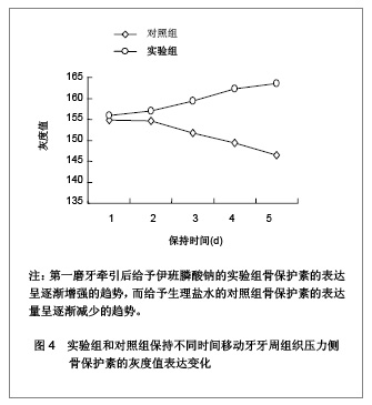
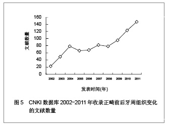
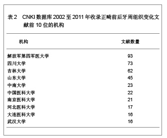
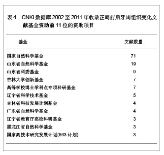
| [1] 刘东刚,王杰军.双膦酸盐类药物的发展[J].中国肿瘤临床,2003, 30(9): 678-683.[2] Pivot X, Lortholary A, Abadie-Lacourtoisie S, et al. Renal safety of ibandronate 6 mg infused over 15 min versus 60 min in breast cancer patients with bone metastases: a randomized open-label equivalence trial. Breast. 2011; 20(6):510-514.[3] Walter C, Al-Nawas B, du Bois A, et al. Incidence of bisphosphonate-associated osteonecrosis of the jaws in breast cancer patients. Cancer. 2009;115(8):1631-1637.[4] Fischer RG, KlingeB. Clinical and histological evaluation of ligature-induced periodontitis in the domestic ferret. J Clin Periodontol. 1994;21(4):230-239.[5] King GJ, Keeling SD, McCoy EA, et al. Measuring dental drift and orthodontic tooth movement in response to various initial forces in adult rat. Am J Orthod Dentofacial Orthop. 1991; 99(5):456-465.[6] 段继强,王宸,张磊,等.骨折局部应用伊班膦酸钠最适剂量选择的动物实验研究[J].现代医学,2010,38(2):96-101.[7] 刘世颖,陈远萍,刘天悦,等.实验性正畸牙移动保持期动物模型的建立[J].中国实用口腔科杂志,2009,2(9):537-539.[8] 郑伟,汪宝军,叶立民.灰度值、光学密度值与免疫组化片阳性表达强弱的关系[J].临床与实验病理学杂志,2003,19(5):566-567.[9] 莫水学,陈扬熙.孕鼠正畸牙移动牙周组织DMP1表达变化的研究[J].广西医科大学学报,2010,27(06):826-828.[10] 王光,丁寅,潘峰,等.大鼠正畸牙移动中牙周组织内STRO-1动态变化的研究[J].口腔医学研究,2008,24(02):124-126.[11] 郭杰,赵青,陈扬熙.动情周期不同阶段正畸牙移动影响牙周组织胰岛素样生长因子表达的研究[J].华西口腔医学杂志,2008, 26(04):439-442.[12] 郭杰,赵青,陈扬熙,等.正畸牙移动对大鼠血清和局部牙周组织雌激素水平的影响[J].上海口腔医学,2007,16(06):618-622.[13] 孙应明.P物质缓释系统对正畸大鼠牙周破骨细胞数目的影响[J].中国美容医学,2007,16(02):153-156.[14] 孙应明,罗颂椒.免疫荧光法观察正畸大鼠牙周P物质神经纤维变化的研究[J].东南国防医药,2006,8(04):249-250,260.[15] 张晓东,林珠,李永明,等.大鼠牙齿移动牙周组织中Ⅰ型胶原和MMP-1及TIMP-1的表达[J].中国美容医学,2005,14(01):12-14.[16] 刘建林,林珠,李永明.局部注射VEGF对大鼠正畸牙齿移动的影响[J].中国美容医学,2004,13(03):272-274.[17] 李永明,罗颂椒.破骨细胞分化因子mRNA在大鼠正畸牙槽骨改建过程中的表达和分布[J].牙体牙髓牙周病学杂志,2004,14(05): 250-253.[18] 常少海,叶剑涛,刘东雄,等.碱性成纤维细胞生长因子在正畸大鼠牙周组织中的表达[J].新医学,2003,34(z1):77-79.[19] Soares PB, Fernandes Neto AJ, Magalhães D, et al. Effect of bone loss simulation and periodontal splinting on bone strain: Periodontal splints and bone strain. Arch Oral Biol. 2011;56 (11): 1373-1381.[20] Lossdörfer S, Abuduwali N, Jäger A. Bone morphogenetic protein-7 modifies the effects of insulin-like growth factors and intermittent parathyroid hormone (1-34) on human periodontal ligament cell physiology in vitro. J Periodontol. 2011;82(6): 900-908.[21] Dao V, Renjen R, Prasad HS, et al. Cementum, pulp, periodontal ligament, and bone response after direct injury with orthodontic anchorage screws: a histomorphologic study in an animal model. J Oral Maxillofac Surg. 2009;67(11): 2440-2445.[22] Murphy KG, Wilcko MT, Wilcko WM, et al. Periodontal accelerated osteogenic orthodontics: a description of the surgical technique. J Oral Maxillofac Surg. 2009;67(10): 2160-2166.[23] Cattaneo PM, Dalstra M, Melsen B. Strains in periodontal ligament and alveolar bone associated with orthodontic tooth movement analyzed by finite element. Orthod Craniofac Res. 2009;12(2):120-128.[24] Cornelis MA, Scheffler NR, Mahy P, et al. Modified miniplates for temporary skeletal anchorage in orthodontics: placement and removal surgeries. J Oral Maxillofac Surg. 2008;66(7): 1439-1445.[25] Sidiropoulou-Chatzigiannis S, Kourtidou M, Tsalikis L. The effect of osteoporosis on periodontal status, alveolar bone and orthodontic tooth movement. A literature review. J Int Acad Periodontol. 2007;9(3):77-84.[26] Bildt MM, Henneman S, Maltha JC, et al. CMT-3 inhibits orthodontic tooth displacement in the rat. Arch Oral Biol. 2007; 52(6):571-578.[27] Meikle MC. Remodeling the dentofacial skeleton: the biological basis of orthodontics and dentofacial orthopedics. J Dent Res. 2007;86(1):12-24.[28] Yoshimatsu M, Shibata Y, Kitaura H, et al. Experimental model of tooth movement by orthodontic force in mice and its application to tumor necrosis factor receptor-deficient mice. J Bone Miner Metab. 2006;24(1):20-27.[29] Risubut S, Teerakapong A, Vattraphodes T, et al. Effect of local delivery of alendronate on bone formation in bioactive glass grafting in rats. Oral Surg Oral Med Oral Pathol Oral Radiol Endod. 2007;104(4):e11-16.[30] Russell RG, Xia Z, Dunford JE, et al. Bisphosphonates:an update on mechanisms of action and how these relate to clinical efficacy. Ann N Y Acad Sci. 2007;1117:209-257.[31] Xiong Y, Yang HJ, Feng J, et al. Effects of alendronate on the proliferation and osteogenic differentiation of MG-63 cells. J Int Med Res. 2009;37(2):407-416.[32] Kim HK, Kim JH, Abbas AA, et al. Alendronate enhances osteogenic differentiation of bone marrow stromal cells:a preliminary study. Clin Orthop Relat Res. 2009;467(12): 3121-3128.[33] Kwon BS, Wang S, Udagawa N, et al. TRI,a new member of the tumor necrosis factor receptor family, induces fibroblast proliferation and inhibits osteoclastogenesis and bone resorption. FASEB J. 1998;12(10):845-854.[34] Udagawa N, Takahashi N, Jimi E, Matsuzaki, et al. Osteoblasts/stromal cells stimulate osteoclast activation through expression of osteoclast differentiation factor RANKL but not macrophage colony-stimulating factor. Bone. 1999; 25(5):517-523. |