中国组织工程研究 ›› 2013, Vol. 17 ›› Issue (6): 1064-1068.doi: 10.3969/j.issn.2095-4344.2013.06.019

• 干细胞培养与分化 stem cell culture and differentiation • 上一篇    下一篇

大鼠骨髓与外周血来源内皮祖细胞生物学特性比较

曹广煜,陈庆伟,李兴升,杨 彦,李桂琼   

  1. 重庆医科大学附属第二医院老年科,重庆市 400010
  • 收稿日期:2012-05-04 修回日期:2012-07-28 出版日期:2013-02-05 发布日期:2013-02-05
  • 作者简介:曹广煜★,男,1984年生,山东省临沂市人,汉族,2012年重庆医科大学毕业,硕士,主要从事干细胞移植治疗缺血性疾病研究。 cgyu9818@163.com

Biological characteristics of rat peripheral blood versus bone marrow derived endothelial progenitor cells

Cao Guang-yu, Chen Qing-wei, Li Xing-sheng, Yang Yan, Li Gui-qiong   

  1. Department of Gerontology, Second Affiliated Hospital of Chongqing Medical University, Chongqing 400010, China
  • Received:2012-05-04 Revised:2012-07-28 Online:2013-02-05 Published:2013-02-05
  • About author:Cao Guang-yu★, Master, Department of Gerontology, Second Affiliated Hospital of Chongqing Medical University, Chongqing 400010, China cgyu9818@163.com

摘要:

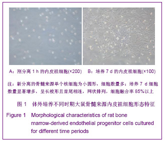
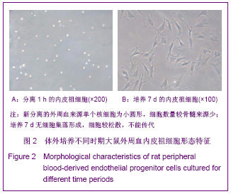
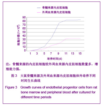
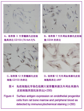
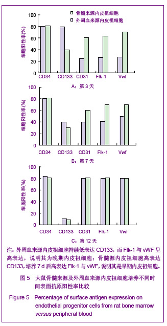
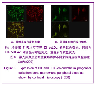
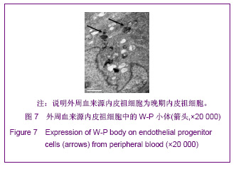
背景:内皮祖细胞不仅参与胚胎血管生成,也参与出生后血管发生和血管内膜损伤后修复,对治疗缺血性疾病意义重大,但目前对内皮祖细胞的分离、培养、鉴定还存在争议。
目的:体外分离、培养大鼠骨髓与外周血来源的内皮祖细胞,并比较其生物学特性。
方法:密度梯度离心法分离SD大鼠骨髓和外周血单个核细胞,接种于纤维连接蛋白铺被的培养瓶中贴壁培养,用加入血管内皮生长因子、碱性成纤维细胞生长因子及表皮生长因子的完全培养基诱导培养,对获得的贴壁细胞进行细胞形态学,免疫细胞化学染色,流式细胞仪,透射电镜,以及Dil-acLDL、FITC-UEA-1双荧光染色法检测。
结果与结论:骨髓来源的内皮祖细胞数量多,集落状生长,增殖能力强;外周血来源的内皮祖细胞数量较少,散在生长,消化后能贴壁但不能传代。两种不同来源的内皮祖细胞免疫细胞化学检测贴壁细胞CD133、CD34、Flk-1、Ⅷ因子在不同时段呈阳性表达;激光共聚焦显微镜观察,Dil-acLDL、FITC-UEA-1均为双染。透射电镜检查外周血来源的内皮祖细胞发现W-P小体。提示大鼠骨髓和外周血均能分离培养出内皮祖细胞,但前者是早期内皮祖细胞,后者为晚期内皮祖细胞,两者生物学特性各不相同。

关键词: 干细胞, 干细胞培养与分化, 内皮祖细胞, 骨髓, 外周血, 分离, 鉴定, 免疫细胞化学染色, 早期内皮祖细胞, 晚期内皮祖细胞, 生物学特性, 大鼠, 干细胞图片文章

Abstract:

BACKGROUND: Endothelial progenitor cells (EPCs) not only participate in embryonic angiogenesis, but also participate in postnatal angiogenesis and vascular intima damage repair, exhibiting a great significance for the treatment of ischemic diseases. Nevertheless, the isolation, culture and identification of endothelial progenitor cells remain controversial.
OBJECTIVE: To isolate and culture endothelial progenitor cells from peripheral blood and bone marrow in vitro, and to compare their biological characteristics.
METHODS: SD rat bone marrow and peripheral blood mononuclear cells were isolated by density gradient centrifugation, inoculated on fibrinin-coated culture flask and cultured with complete medium containing vascular endothelial growth factor, basic fibroblast growth, and epidermal growth factor. Cell morphplogy, immunocytochemical staining, flow cytometry and transmission electron microscopy as well as Dil-acLDL, FITC-UEA-1 double fluorescence staining were performed on the harvested adherent cells.
RESULTS AND CONCLUSION: A large number of endothelial progenitor cells were harvested from bone marrow, and they were grown in a colony-like manner and show strong proliferative capacity. A small number of endothelial progenitor cells were harvested from peripheral blood. They were grown in a sparse manner and could adhere to flask wall but could not be passaged. Immunocytochemical staining showed that adherent cells from two sources were positive for CD133, CD34, Flk-1 and VII factors at different time periods. Laser confocal microscopy showed that adherent cells from two sources can stain Dil-acLDL and FITC-UEA-1. Transmission electron microscopy showed that W-P body was found in the peripheral blood endothelial progenitor cells. These results indicate that endothelial progenitor cells can be cultured from both bone marrow and peripheral blood, but early-stage endothelial progenitor cells can be cultured from bone marrow and late-stage endothelial progenitor cells can be cultured from peripheral blood. These two kinds of cells show different biological characteristics.

Key words: stem cells, stem cell culture and differentiation, endothelial progenitor cells, bone marrow, peripheral blood, isolation, identification, immunocytochemical staining, early endothelial progenitor cell, late-stage endothelial progenitor cells, biological characteristics, rats, stem cell photographs-containing paper

中图分类号: