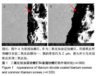
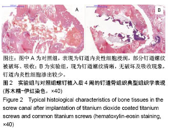
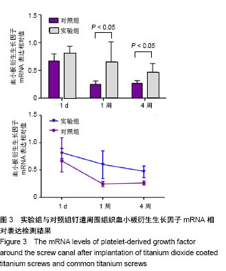
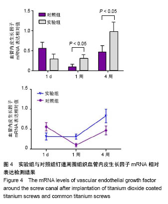
| [1] Moroni A, Vannini F, Mosca M, et al.State of the art review: techniques to avoid pin loosening and infection in external fixation. J Orthop Trauma.2002;16(3):189-195.[2] Mutsuzaki H, Sogo Y, Oyane A, et al.Improved bonding of partially osteomyelitic bone to titanium pins owing to biomimetic coating of apatite.Int J Mol Sci. 2013;14(12): 24366-14379.[3] Pieske O, Kaltenhauser F, Pichlmaier L, et al.Clinical benefit of hydroxyapatite-coated pins compared with stainless steel pins in external fixation at the wrist: A randomised prospective study. Injury. 2010;41(10):1031-1036.[4] Holt J, Hertzberg B, Weinhold P, et al.Decreasing bacterial colonization of external fixation pins through nitric oxide release coatings.J Orthop Trauma. 2011;25(7):432-437.[5] Guo C, Wang K, Hou S, et al. H2O2 and/or TiO2 photocatalysis under UV irradiation for the removal of antibiotic resistant bacteria and their antibiotic resistance genes. J Hazard Mater. 2017; 323(Pt B):710-718.[6] Jimenez-Tototzintle M, Ferreira IJ, Da Silva Duque S, et al. Removal of contaminants of emerging concern (CECs) and antibiotic resistant bacteria in urban wastewater using UVA/TiO2/H2O2 photocatalysis. Chemosphere. 2018;210: 449-457.[7] Moreira NFF, Narciso-Da-Rocha C, Polo-Lopez MI, et al.Solar treatment (H2O2, TiO2-P25 and GO-TiO2 photocatalysis, photo-Fenton) of organic micropollutants, human pathogen indicators, antibiotic resistant bacteria and related genes in urban wastewater. Water Res.2018;135:195-206.[8] Esfandiari N, Simchi A, Bagheri R.Size tuning of Ag-decorated TiO(2) nanotube arrays for improved bactericidal capacity of orthopedic implants. J Biomed Mater Res A.2014;102(8):2625-2635.[9] Zhang H,Sun Y,Tian A,et al.Improved antibacterial activity and biocompatibility on vancomycin-loaded TiO2 nanotubes: in vivo and in vitro studies. Int J Nanomedicine. 2013;8: 4379-4389.[10] Maurer TB, Ochsner PE, Schwarzer G, et al.Increased loosening of cemented straight stem prostheses made from titanium alloys. An analysis and comparison with prostheses made of cobalt-chromium-nickel alloy.Int Orthop. 2001;25(2): 77-80.[11] Shim J, Seo YS, Oh BT, et al. Microbial inactivation kinetics and mechanisms of carbon-doped TiO 2 (C-TiO 2) under visible light. J Hazard Mater. 2016;306:133-139.[12] Gao A, Hang R, Huang X, et al.The effects of titania nanotubes with embedded silver oxide nanoparticles on bacteria and osteoblasts. Biomaterials. 2014;35(13): 4223-4235.[13] Leitner L, Malaj I, Sadoghi P, et al.Pedicle screw loosening is correlated to chronic subclinical deep implant infection: a retrospective database analysis. Eur Spine J. 2018; 27(10): 2529-2535.[14] Agrawal A. Pedicle screw nut loosening: potentially avoidable causes of spine instrumentation failure. Asian Spine J. 2014; 8(2): 224-226.[15] Lawes TJ, Scott JC, Goodship AE.Increased insertion torque delays pin-bone interface loosening in external fixation with tapered bone screws. J Orthop Trauma.2004;18(9):617-622.[16] Hee HT, Khan MS, Goh JC, et al.Insertion torque profile during pedicle screw insertion of the thoracic spine with and without violation of the pedicle wall: comparison between cylindrical and conical designs. Spine (Phila Pa 1976). 2006; 31(22):E840-846.[17] Chatzistergos PE, Magnissalis EA, Kourkoulis SK.Numerical simulation of bone screw induced pretension: the cases of under-tapping and conical profile. Med Eng Phys. 2014;36(3): 378-386.[18] Pettine KA, Chao EY, Kelly PJ. Analysis of the external fixator pin-bone interface. Clin Orthop Relat Res. 1993;(293):18-27.[19] Siamos G, Winkler S, Boberick KG. Relationship between implant preload and screw loosening on implant-supported prostheses.J Oral Implantol.2002;28(2): 67-73.[20] 姚珍松,唐永超,陈康,等.骨水泥螺钉强化固定伴骨质疏松腰椎滑脱症的稳定性及椎间融合[J].中国组织工程研究, 2016,20(4): 517-521.[21] Partale K, Klein P, Schell H, et al.Poly(D,L-lactide) coating is capable of enhancing osseous integration of Schanz screws in the absence of infection. J Biomed Mater Res B Appl Biomater. 2005; 74(1):608-616.[22] Rammelt S, Illert T, Bierbaum S, et al.Coating of titanium implants with collagen, RGD peptide and chondroitin sulfate. Biomaterials.2006;27(32):5561-5571.[23] 韩建业,于振涛,周廉.羟基磷灰石/TiO2生物活性复合涂层的研究进展[C].第六届中国功能材料及其应用学术会议论文集, 2007:1808-1812.[24] Brammer KS, Oh S, Cobb CJ, et al.Improved bone-forming functionality on diameter-controlled TiO2 nanotube surface. Acta Biomaterialia. 2009;5(8):3215-3223.[25] Ercan B, Webster TJ. The effect of biphasic electrical stimulation on osteoblast function at anodized nanotubular titanium surfaces. Biomaterials. 2010;31(13):3684-3693.[26] Uhm SH, Song DH, Kwon JS, et al.Tailoring of antibacterial Ag nanostructures on TiO2 nanotube layers by magnetron sputtering.J Biomed Mater Res B Appl Biomater. 2014; 102(3):592-603.[27] Singh AV, Vyas V, Patil R, et al. Quantitative characterization of the influence of the nanoscale morphology of nanostructured surfaces on bacterial adhesion and biofilm formation. PLoS One.2011;6(9): e25029.[28] Gunasekaran T, Nigusse T, Dhanaraju MD. Silver nanoparticles as real topical bullets for wound healing. J Am Coll Clin Wound Spec. 2011;3(4):82-96.[29] Franchini A, Ottaviani E. Repair of molluscan tissue injury: role of PDGF and TGF-beta1. Tissue Cell. 2000;32(4): 312-321.[30] Si HP, Lu ZH, Lin YL, et al. Transfect bone marrow stromal cells with pcDNA3.1-VEGF to construct tissue engineered bone in defect repair. Chin Med J (Engl). 2012;125(5): 906-911.[31] Eming SA, Krieg T. Molecular mechanisms of VEGF-A action during tissue repair. J Investig Dermatol Symp Proc. 2006; 11(1):79-86.[32] Hong YK, Lange-Asschenfeldt B,Velasco P, et al. VEGF-A promotes tissue repair-associated lymphatic vessel formation via VEGFR-2 and the alpha1beta1 and alpha2beta1 integrins. FASEB J. 2004; 18(10): 1111-1113.[33] Mutsuzaki H,Ito A,Sakane M,et al. Fibroblast growth factor-2-apatite composite layers on titanium screw to reduce pin tract infection rate. J Biomed Mater Res B Appl Biomater. 2008;86(2):365-374.[34] Mutsuzaki H, Ito A, Sakane M, et al. Calcium phosphate coating formed in infusion fluid mixture to enhance fixation strength of titanium screws. J Mater Sci Mater Med. 2007; 18(9):1799-1808. |
.jpg)
.jpg)