中国组织工程研究 ›› 2019, Vol. 23 ›› Issue (19): 2959-2964.doi: 10.3969/j.issn.2095-4344.1240
• 骨组织构建 bone tissue construction • 上一篇 下一篇
溶血磷脂酸刺激可影响成骨细胞表达结缔组织生长因子
胡 广,张开伟,徐远坤
- (贵阳中医学院第一附属医院骨科,贵州省贵阳市 550001)
Lysophosphatidic acid affects the expression of connective tissue growth factor in osteoblasts
Hu Guang, Zhang Kaiwei, Xu Yuankun
- (Department of Orthopedics, the First Affiliated Hospital of Guiyang University of Chinese Medicine, Guiyang 550001, Guizhou Province, China)
摘要:
文章快速阅读:
.jpg) 文题释义:
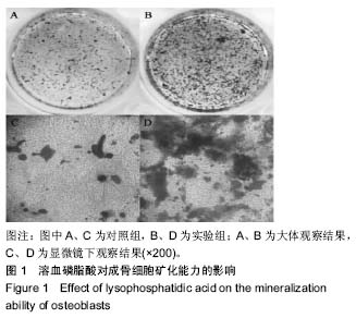
溶血磷脂酸:是一种具有多种生物活性的磷脂,在人体内广泛存在,可参与一系列的生物学过程,包括细胞增殖、分化、凋亡、生存、趋化和肿瘤细胞侵袭等。骨折后局部形成血肿,血肿中活化的血小板可释放出大量的溶血磷脂酸,这些局部聚集的溶血磷脂酸可能参与骨折的愈合过程,但其在骨折中所起的作用仍不清楚。体外研究证实溶血磷脂酸可诱导间充质干细胞向成骨细胞向分化,并促进其形成矿化结节的能力。
结缔组织生长因子2:是富含半胱氨酸的多肽,是CCN蛋白家族中的一员,是强力的促结缔组织增生的生长因子,最初的研究发现其参与多种包括细胞增殖、迁移、黏附和生存、分化及细胞外基质合成。大量的研究表明其与多种器官的纤维化密切相关,在多种组织内广泛表达,并在发育中起主要作用,包括骨组织的发育和形成。
文题释义:
溶血磷脂酸:是一种具有多种生物活性的磷脂,在人体内广泛存在,可参与一系列的生物学过程,包括细胞增殖、分化、凋亡、生存、趋化和肿瘤细胞侵袭等。骨折后局部形成血肿,血肿中活化的血小板可释放出大量的溶血磷脂酸,这些局部聚集的溶血磷脂酸可能参与骨折的愈合过程,但其在骨折中所起的作用仍不清楚。体外研究证实溶血磷脂酸可诱导间充质干细胞向成骨细胞向分化,并促进其形成矿化结节的能力。
结缔组织生长因子2:是富含半胱氨酸的多肽,是CCN蛋白家族中的一员,是强力的促结缔组织增生的生长因子,最初的研究发现其参与多种包括细胞增殖、迁移、黏附和生存、分化及细胞外基质合成。大量的研究表明其与多种器官的纤维化密切相关,在多种组织内广泛表达,并在发育中起主要作用,包括骨组织的发育和形成。摘要
背景:溶血磷脂酸可刺激上皮细胞和成肌细胞表达结缔组织生长因子2,但涉及的分子机制至今仍不清楚。成骨细胞是骨发育和骨折愈合中的重要细胞,其也表达溶血磷脂酸的受体,骨折局部高表达溶血磷脂酸和成骨细胞,且结缔组织生长因子2在骨折愈合中发挥主要作用。
目的:研究外源性溶血性磷脂酸对成骨细胞中结缔组织生长因子2表达水平的影响及其分子机制。
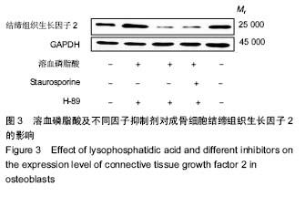
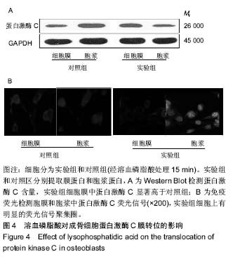
方法:①体外培养MC3T3-E1细胞,将细胞分为对照组和实验组,对照组仅给予矿化诱导液,实验组在矿化诱导液中加入终浓度为20 μmol/L的溶血磷脂酸,刺激0.5,1,2,4,6,8及12 h,提取RNA并检测结缔组织生长因子2 mRNA表达水平;②MC3T3-E1细胞预先经蛋白激酶A抑制剂(H-89)和蛋白激酶C抑制剂(Staurosporine)处理0.5 h,再加入溶血磷脂酸。定量PCR检测结缔组织生长因子2 mRNA表达水平、Western Blot法检测结缔组织生长因子2蛋白表达水平,Western blot法和免疫荧光检测细胞蛋白激酶C转位情况。
结果与结论:①溶血磷脂酸作用于MC3T3-E1细胞后,可迅速引起细胞内结缔组织生长因子2 mRNA和蛋白表达水平提高,刺激后2 h其表达量达到峰值,随后表达水平随时间逐渐降低,至8 h和12 h已基本恢复至刺激前的表达水平;②Staurosporine可明显减弱溶血磷脂酸刺激成骨细胞表达结缔组织生长因子2的能力;而H-89则可以使溶血磷脂酸刺激成骨细胞表达结缔组织生长因子2的能力进一步增强;③成骨细胞经溶血磷脂酸刺激15 min后,溶血磷脂酸可明显提高细胞膜蛋白激酶C表达水平,同时胞浆中蛋白激酶C表达水平降低;④结果提示,溶血磷脂酸可刺激成骨细胞表达结缔组织生长因子2,这一过程可能涉及蛋白激酶A和蛋白激酶C途径。
中国组织工程研究杂志出版内容重点:组织构建;骨细胞;软骨细胞;细胞培养;成纤维细胞;血管内皮细胞;骨质疏松;组织工程
ORCID: 0000-0002-4803-2242(胡广)
中图分类号:

.jpg) 文题释义:
溶血磷脂酸:是一种具有多种生物活性的磷脂,在人体内广泛存在,可参与一系列的生物学过程,包括细胞增殖、分化、凋亡、生存、趋化和肿瘤细胞侵袭等。骨折后局部形成血肿,血肿中活化的血小板可释放出大量的溶血磷脂酸,这些局部聚集的溶血磷脂酸可能参与骨折的愈合过程,但其在骨折中所起的作用仍不清楚。体外研究证实溶血磷脂酸可诱导间充质干细胞向成骨细胞向分化,并促进其形成矿化结节的能力。
结缔组织生长因子2:是富含半胱氨酸的多肽,是CCN蛋白家族中的一员,是强力的促结缔组织增生的生长因子,最初的研究发现其参与多种包括细胞增殖、迁移、黏附和生存、分化及细胞外基质合成。大量的研究表明其与多种器官的纤维化密切相关,在多种组织内广泛表达,并在发育中起主要作用,包括骨组织的发育和形成。
文题释义:
溶血磷脂酸:是一种具有多种生物活性的磷脂,在人体内广泛存在,可参与一系列的生物学过程,包括细胞增殖、分化、凋亡、生存、趋化和肿瘤细胞侵袭等。骨折后局部形成血肿,血肿中活化的血小板可释放出大量的溶血磷脂酸,这些局部聚集的溶血磷脂酸可能参与骨折的愈合过程,但其在骨折中所起的作用仍不清楚。体外研究证实溶血磷脂酸可诱导间充质干细胞向成骨细胞向分化,并促进其形成矿化结节的能力。
结缔组织生长因子2:是富含半胱氨酸的多肽,是CCN蛋白家族中的一员,是强力的促结缔组织增生的生长因子,最初的研究发现其参与多种包括细胞增殖、迁移、黏附和生存、分化及细胞外基质合成。大量的研究表明其与多种器官的纤维化密切相关,在多种组织内广泛表达,并在发育中起主要作用,包括骨组织的发育和形成。