中国组织工程研究 ›› 2019, Vol. 23 ›› Issue (7): 1057-1062.doi: 10.3969/j.issn.2095-4344.1023

• 组织构建实验造模 experimental modeling in tissue construction • 上一篇    下一篇

野生型小鼠慢性根尖周炎模型PLCγ2的表达

杨淳贺,仉  红,董  明,王丽娜,牛卫东   

  1. (大连医科大学口腔医学院牙体牙髓教研室,辽宁省大连市  116041)
  • 收稿日期:2018-08-23 出版日期:2019-03-08 发布日期:2019-03-08
  • 作者简介:杨淳贺,女,1990年生,辽宁省辽阳市人,汉族,大连医科大学在读硕士,主要从事牙体牙髓疾病的研究。
  • 基金资助:

    国家自然科学基金(81171538),项目负责人:牛卫东

Expression of PLCgamma2 in a wild-type mouse model of chronic periapical periodontitis

Yang Chunhe, Zhang Hong, Dong Ming, Wang Lina, Niu Weidong   

  1.  (Department of Endodontology, School of Stomatology, Dalian Medical University, Dalian 116041, Liaoning Province, China)
  • Received:2018-08-23 Online:2019-03-08 Published:2019-03-08
  • About author:Yang Chunhe, Master candidate, Department of Endodontology, School of Stomatology, Dalian Medical University, Dalian 116041, Liaoning Province, China
  • Supported by:

    the National Natural Science Foundation of China, No. 81171538 (to NWD)

摘要:

文章快速阅读:

文题释义:
PLCγ2:全称Phospholipase Cγ2,是一种磷脂酶,参与了生物体的许多生理病理过程,包括细胞增殖和分化、调节细胞骨架、细胞凋亡、肿瘤的迁移,同时其在免疫细胞和破骨细胞的形成过程中也发挥了不同的作用。PLCγ2通过一系列反应激活钙离子(Ca2+)信号通路,调节破骨细胞的生成。
慢性根尖周炎:是由多种细菌及毒力因子入侵根尖周组织,引起根尖组织炎症性反应,以根尖区的牙槽骨吸收为主要特征的一类疾病,是多种细胞因子共同参与的结果。
摘要
背景
:研究表明,抑制BTK-PLCγ2- Ca2+信号通路,破骨细胞的形成和功能受到抑制,说明PLCγ2参与了破骨细胞的形成和分化。
目的:通过建立野生型小鼠慢性根尖周炎模型,观察PLCγ2在小鼠实验性根尖周炎组织中的表达。
方法:选用20只C57BL/6L野生型雄性小鼠,随机选取16只为实验组行双侧下颌第1磨牙开髓术,并将开髓后的髓腔暴露于口腔环境,诱导形成根尖周炎病损。分别于开髓后1,2,3和4周各随机处死4只小鼠,分离下颌骨。剩余4只不开髓作为空白对照,0周时处死后分离下颌骨。制作冰冻切片后用苏木精-伊红染色方法观察小鼠根尖组织炎症情况,免疫组织化学染色检测PLCγ2的表达与分布情况,酶组织化学染色检测根尖组织炎症区域破骨细胞的表达。
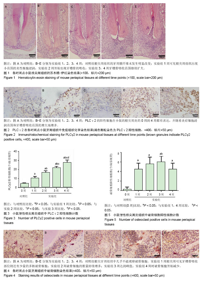
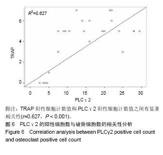
结果与结论:①苏木精-伊红染色结果:对照组小鼠下颌第1磨牙牙周组织几乎无炎性细胞浸润;实验组1-4周,下颌第1磨牙牙周组织炎性细胞浸润范围越来越大,牙槽骨破坏也逐渐增多,说明成功建立了小鼠根尖周炎模型;②免疫组织化学染色结果:发现PLCγ2在小鼠根尖周炎的0-4周都有表达;对照组根尖周组织中仅有少量PLCγ2的表达;实验组1-4周PLCγ2阳性表达随炎症细胞浸润范围的逐渐扩大而增长(P < 0.05);③TRAP染色结果:对照组根尖牙周组织中几乎不能观察到破骨细胞;实验组1周,可观察到少量的多核破骨细胞;实验组2周,破骨细胞的数量持续增多;实验组3周达到峰值;实验组4周破骨细胞数量逐渐减少;与对照组相比差异均有显著性意义(P < 0.05);④相关性分析:TRAP阳性细胞计数值和PLCγ2阳性细胞计数之间有显著相关性(r=0.627,P < 0.001);⑤结果说明,PLCγ2在小鼠根尖周组织中有表达,提示其可能与根尖周炎症反应和骨吸收作用相关。

中国组织工程研究杂志出版内容重点:组织构建;骨细胞;软骨细胞;细胞培养;成纤维细胞;血管内皮细胞;骨质疏松组织工程
ORCID: 0000-0002-8786-7656(杨淳贺)

关键词: PLCγ2, 尖周炎, 小鼠慢性根尖周炎模型, 破骨细胞, 组织构建

Abstract:

BACKGROUND: Inhibiting BTK-PLCγ2-Ca2+ signaling pathway has been shown to inhibit formation and function of osteoclasts, suggesting that PLCγ2 participates in cell formation and differentiation.
OBJECTIVE: To study the expression of PLCγ2 in periapical tissues by establishing a model of periapical periodontitis in wild-type mice.
METHODS: Twenty male wild-type C57BL/6L mice were selected. Bilateral mandibular first molar pulp cavities of 16 mice were exposed to the oral environment and the formation of apical periodontitis was induced (experimental group). The mice were randomly sacrificed at 1, 2, 3 and 4 weeks and the lower jaws were isolated. The remaining four mice were used as blank control group, and were sacrificed at 0 week to isolate the lower jaws. After being histologically processed, they were hematoxylin-eosin stained to observe the inflammation conditions in the apical area. Expression and distribution of PLCγ2 were analyzed by immunohistochemical staining. Enzyme-histochemical staining was used to detect the expression of osteoclasts in apical inflammatory tissue.
RESULTS AND CONCLUSION: According to the histological examination, there was almost no inflammatory cell infiltration in the periodontal tissue of the mandibular first molar in the blank control group. In the experimental group, at 1 to 4 weeks after operation, the infiltration of inflammatory cells in the periodontal tissues and the destruction of alveolar bone of mandibular first molar increased gradually, indicating that the mouse periapical periodontitis model was established successfully. Immunohistochemical analysis found that the expression of PLCγ2 was detected at 0 to 4 weeks after periapical periodontitis in mice. A small amount of PLCγ2 expressed in normal dental apical tissues. PLCγ2 in the experimental group was mainly expressed in the nucleus of lymphocytes, plasma cells and other cells, and there was a small amount of expression in the cell membrane and cytoplasm. At the1st, 2nd, 3rd, 4th weeks, the positive expression of PLCγ2 also increased due to the gradual expansion of inflammatory cell infiltration. The positive cell counts were all significantly different (P < 0.05). The results of TRAP staining showed that almost no osteoclasts were observed in periapical periodontal tissues of blank control group. In the experimental group, at 1 week after operation, a small amount of multinucleated osteoclasts were observed. The number of osteoclasts continued to increase at 2 weeks after operation. At 3 weeks, the number of osteoclasts reached the peak. At 4 weeks postoperatively, a decrease in the number of osteoclasts was observed. The difference was significant between experimental and control groups (P < 0.05). Correlation analysis between TRAP positive cell count and PLCγ2 positive cell count showed a significant correlation (r=0.627, P < 0.001). These results indicate that PLCγ2 is expressed in periapical tissues of mice, implying that it may be related to periapical inflammatory reaction and bone resorption.

中国组织工程研究杂志出版内容重点:组织构建;骨细胞;软骨细胞;细胞培养;成纤维细胞;血管内皮细胞;骨质疏松组织工程

Key words: Phospholipase C gamma, Periapical Periodontitis, Osteoclasts, Tissue Engineering

中图分类号: