中国组织工程研究 ›› 2018, Vol. 22 ›› Issue (32): 5157-5162.doi: 10.3969/j.issn.2095-4344.0539

• 软骨组织构建 cartilage tissue construction • 上一篇    下一篇

姜黄素对脂多糖诱导炎症致软骨细胞凋亡及增殖能力的影响

李亚楠,倪  娟,方禹舜,李  涛,谈鸿飞,张青松   

  1. 华中科技大学同济医学院附属普爱医院,湖北省武汉市  430030
  • 收稿日期:2018-05-20 出版日期:2018-11-18 发布日期:2018-11-18
  • 通讯作者: 张青松,硕士生导师,副主任医师,华中科技大学同济医学院附属普爱医院, 湖北省武汉市 430030
  • 作者简介:李亚楠,男,1989年生,河南省驻马店市人,汉族,2014年华中科技大学同济医学院毕业,博士,医师,主要从事关节退行性疾病、软骨及韧带损伤研究。
  • 基金资助:

    武汉市卫生计生委科研计划资助项目(WZ18E07)

Effects of curcumin on apoptosis and proliferation of inflammatory chondrocytes induced by lipopolysaccharide

Li Ya-nan, Ni Juan, Fang Yu-shun, Li Tao, Tan Hong-fei, Zhang Qing-song   

  1. Puai Hospital Affiliated to Tongji Medical College of Huazhong University of Science and Technology, Wuhan 430030, Hubei Province, China
  • Received:2018-05-20 Online:2018-11-18 Published:2018-11-18
  • Contact: Zhang Qing-song, Master’s supervisor, Associate chief physician, Puai Hospital Affiliated to Tongji Medical College of Huazhong University of Science and Technology, Wuhan 430030, Hubei Province, China
  • About author:Li Ya-nan, MD, Physician, Puai Hospital Affiliated to Tongji Medical College of Huazhong University of Science and Technology,Wuhan 430030, Hubei Province, China
  • Supported by:

    the Scientific Research Program of Health and Family Planning Commission of Wuhan Municipality, No. WZ18E07

摘要:

文章快速阅读:

文题释义:
姜黄素:是一种从姜科植物姜黄等的根茎中提取得到的黄色色素,为酸性多酚类物质,主链为不饱和脂族及芳香族基团。现代研究发现姜黄素可以抑制炎症反应、抗氧化、抗类风湿、降脂、抗动脉粥样硬化、抗衰老消除自由基及抑制肿瘤生长的作用。
软骨细胞:软骨组织由软骨细胞和细胞外基质构成,软骨细胞是软骨组织中唯一的细胞成分,是关节软骨破坏过程中的靶细胞,因此研究靶细胞的变化是探索关节软骨病变和治疗的重要方法。软骨细胞炎症模型是指采用脂多糖体外刺激培养的软骨细胞诱发炎症反应,是研究骨关节炎良好的体外模型。
摘要
背景
:在骨关节炎的相关研究中,姜黄素被证实具有抗炎抗氧化及抗凋亡的作用,但相关机制的研究还不够深入。
目的:探讨姜黄素对脂多糖诱导的软骨细胞凋亡、增殖的影响,以及潜在的作用机制,为治疗骨关节炎疾病提供新思路。
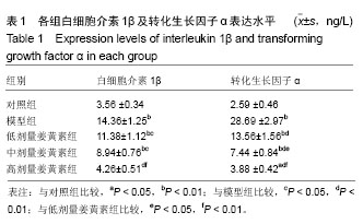
方法:取4周龄SD大鼠,进行软骨细胞的体外分离培养,甲苯胺蓝染色进行细胞鉴定。采用脂多糖诱导法建立体外软骨细胞炎症模型。将软骨细胞分为5组:对照组(普通培养基);模型组(含5 µg/L脂多糖培养基);低剂量姜黄素组(含5 µg/L脂多糖+5 µmol/L姜黄素培养基);中剂量姜黄素组(含5 µg/L脂多糖+10 µmol/L姜黄素培养基);高剂量姜黄素组(含5 µg/L脂多糖+20 µmol/L姜黄素培养基)。培养24 h后,ELISA法检测各组细胞上清液中白细胞介素1β和转化生长因子α的水平;流式细胞仪检测细胞凋亡状况;MTT法进行细胞增殖检测;Western blot法检测自噬相关蛋白及ERK1/2的表达变化。
结果与结论:①经脂多糖诱导处理的软骨细胞上清液中白细胞介素1β和转化生长因子α的水平均显著高于对照组(P < 0.01),各姜黄素组细胞上清液中白细胞介素1β和转化生长因子α的水平较模型组显著降低(P < 0.05,P < 0.01),且呈剂量依赖性; ②经24 h培养后,模型组细胞凋亡率显著高于对照组(P < 0.01),与模型组相比,各姜黄素处理的细胞凋亡率有所降低,且20 µmol/L姜黄素处理的细胞凋亡率降低最为显著  (P < 0.01);③MTT法检测结果显示,与对照组相比,模型组细胞增殖能力显著降低(P < 0.01);与模型组相比,经不同浓度的姜黄素处理细胞后,细胞的增殖能力得以恢复,且20 µmol/L姜黄素处理的细胞恢复效果最好;④Western blot法检测结果显示,与模型组相比,各姜黄素组细胞自噬能力明显增强,自噬标志物LC3-II及相关蛋白Beclin-1表达量均显著升高(P < 0.05,P < 0.01),且p-ERK1/2蛋白表达量显著升高(P < 0.05);⑤结果提示,20 µmol/L姜黄素能显著缓解脂多糖诱导的软骨细胞炎症反应,抑制软骨细胞凋亡,并对脂多糖诱导的炎症软骨细胞增殖抑制具有一定的拮抗作用,这可能与姜黄素通过ERK1/2途径激活的自噬分子调控紧密相关。

中国组织工程研究杂志出版内容重点:组织构建;骨细胞;软骨细胞;细胞培养;成纤维细胞;血管内皮细胞;骨质疏松组织工程
ORCID: 0000-0002-4583-6958(李亚楠)

关键词: 姜黄素, 脂多糖, 软骨细胞, 自噬, 细胞外信号调节激酶1/2, 炎症因子, 骨关节炎, 细胞凋亡, 细胞增殖

Abstract:

BACKGROUND: Curcumin has been shown to play antioxidative, anti-inflammatory and anti-apoptotic roles in osteoarthritis, but the underlying mechanisms remain unclear.
OBJECTIVE: To investigate the effects of curcumin on apoptosis and proliferation of chondrocytes induced by lipopolysaccharide, and to explore the potential mechanisms, so as to provide new ideas for the treatment of osteoarthritis.
METHODS: Chondrocytes were isolated from the 4-week-old Sprague-Dawley rats, cultured, and identified by toluidine blue staining. The chondrocyte inflammation model in vitro was established by lipopolysaccharide induction. The cells were divided into five groups: control group (common medium); model group (medium with 5 µg/L lipopolysaccharide); low-dose group (medium with 5 µg/L lipopolysaccharide +  5 µmol/L curcumin); medium-dose group (medium with 5 µg/L lipopolysaccharide + 10 µmol/L curcumin); high-dose group (medium with     5 µg/L lipopolysaccharide + 20 µmol/L curcumin). After 24 hours, the expression levels of interleukin 1β and transforming growth factor α in the cellular supernatant were detected by ELISA method. The apoptotic cells were detected by flow cytometry. Cell proliferation was tested by MTT assay. The expression levels of autophagy-related proteins and extracellular regulated protein kinases 1/2 were determined by western blot assay.
RESULTS AND CONCLUSION: The levels of interleukin 1β and transforming growth factor α in the supernatant of chondrocytes induced by lipopolysaccharide were significantly higher than those in the control group (P < 0.01). The levels of interleukin 1β and transforming growth factor α in the supernatant treated with curcumin were lower than those in the model group, which was on a dose-dependent manner (P < 0.05, P < 0.01). After 24 hours of treatment, apoptotic rate of cells in the model group was significantly higher than that in the control group (P < 0.01). Compared with the model group, the cell apoptotic rate in each curcumin group was decreased, especially the high-dose group (P < 0.01). MTT assay showed that the cell proliferation ability in the model group was significantly lower than that in the control group (P < 0.01). Compared with the model group, the cell proliferation ability in each curcumin group was restored, and the effect was the best in the high-dose group. The results of western blot showed that the autophagic ability and the levels of autophagy marker (LC3-II) and autophagy related protein (Beclin-1) were significantly increased in each curcumin group compared with the model group(P < 0.05, P < 0.01), and the expression level of p-ERK1/2 protein was significantly increased (P < 0.05). Our findings suggest that 20 µmol/L curcumin cannot only inhibit apoptosis and relieve inflammatory of chondrocytes induced by lipopolysaccharide, but also has a certain antagonistic effect on lipopolysaccharide- induced inhibition of inflammatory chondrocyte proliferation, which may be via regulating the ERK1/2 pathway-activated autophagy.

中国组织工程研究杂志出版内容重点:组织构建;骨细胞;软骨细胞;细胞培养;成纤维细胞;血管内皮细胞;骨质疏松组织工程

Key words: Osteoarthritis, Curcumin, Cell Proliferation, Apoptosis, Tissue Engineering

中图分类号: