中国组织工程研究 ›› 2023, Vol. 27 ›› Issue (26): 4120-4125.doi: 10.12307/2023.450

• 血管组织构建 vascular tissue construction • 上一篇    下一篇

脑源性神经营养因子干预腰椎管狭窄症大鼠神经元活性、疼痛及相关因子的变化

安河朋,刘振腾,李立新,许雅芳,樊国峰   

  1. 河北中石油中心医院骨科,河北省廊坊市  065000
  • 收稿日期:2022-01-17 接受日期:2022-07-09 出版日期:2023-09-18 发布日期:2023-01-20
  • 通讯作者: 樊国峰,硕士,主任医师,河北中石油中心医院骨科,河北省廊坊市 065000
  • 作者简介:安河朋,男,1982年生,河北省大城县人,汉族,2008年河北北方学院毕业,硕士,主治医师,主要从事脊柱外科方向的研究。
  • 基金资助:
    廊坊市科技计划项目(2021013147),项目负责人:安河朋

Effects of brain-derived neurotrophic factor on neuronal activity, pain, and related cytokines in rats with lumbar spinal stenosis

An Hepeng, Liu Zhenteng, Li Lixin, Xu Yafang, Fan Guofeng   

  1. Department of Orthopedics, Hebei Central Hospital of Petro China, Langfang 065000, Hebei Province, China
  • Received:2022-01-17 Accepted:2022-07-09 Online:2023-09-18 Published:2023-01-20
  • Contact: Fan Guofeng, Master, Chief physician, Department of Orthopedics, Hebei Central Hospital of Petro China, Langfang 065000, Hebei Province, China
  • About author:An Hepeng, Master, Attending physician, Department of Orthopedics, Hebei Central Hospital of Petro China, Langfang 065000, Hebei Province, China
  • Supported by:
    Langfang Science and Technology Plan Project, No. 2021013147 (to AHP)

摘要:

文题释义:

缺氧诱导因子1α(hypoxia-inducible factor-1α,HIF-1α):是一种转录因子,主要在缺氧条件下表达,其可激活多种因子的转录,从而提高组织和细胞对缺血、缺氧环境的适应。而有研究表明,脊髓创伤后继发性病理损害常引起脊髓组织缺血、缺氧,故可推测缺氧诱导因子1α可能与腰椎管狭窄症的发生、发展密切相关。
脑源性神经营养因子(brain-derived neurotrophic factor,BDNF):作为神经营养因子家族的重要成员,在神经保护、促进突触生长等方面发挥着重要作用,且目前已有研究报道其在治疗脊髓损伤等神经性疾病的研究中,对周围神经的作用取得了一定的成果,具有较好的临床疗效。

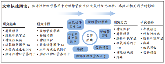
背景:研究发现,脑源性神经营养因子缺失会导致中枢运动结构的神经退行性病变,从而导致多种运动神经疾病的发生,而腰椎管狭窄症作为慢性进行性神经功能障碍症候群,脑源性神经营养因子可能成为治疗该病的有效靶点。
目的:探究脑源性神经营养因子对腰椎管狭窄症大鼠神经元活性、疼痛及缺氧诱导因子1α/血管内皮生长因子的影响。
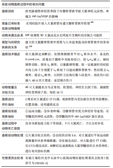
方法:选取40只SPF级SD雄性大鼠,随机取30只建立腰椎管狭窄模型,剩余10只为正常组。建模成功后将大鼠随机分为模型组、神经生长因子组及脑源性神经营养因子组。神经生长因子组大鼠腹腔注射1 500 U的神经生长因子,脑源性神经营养因子组大鼠鞘内注射20 μL 10 mg/L的脑源性神经营养因子,均1次/d,持续30 d;正常组、模型组同期灌胃同体积生理盐水。给药结束后,观察并记录大鼠运动功能及体表疼痛情况,CT检测腰椎管密度,神经干动作点位传导速度测定仪检测大鼠神经传导速度,TUNEL法检测脊髓组织神经元活性,免疫印迹法检测缺氧诱导因子1α/血管内皮生长因子蛋白表达。

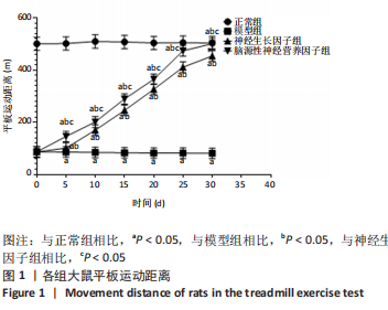
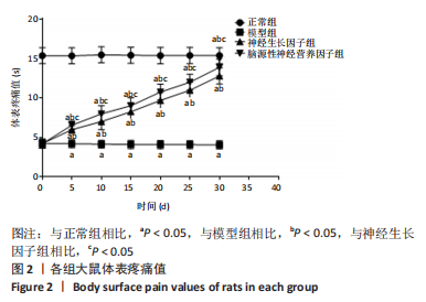
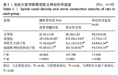
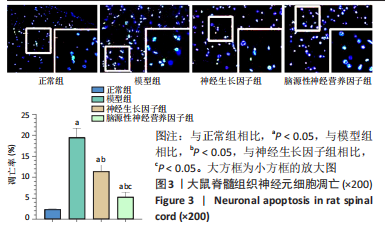
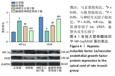
结果与结论:①与正常组比较,模型组大鼠平板运动距离、体表疼痛值、神经传导速度均明显降低(P < 0.05);与模型组比较,神经生长因子组、脑源性神经营养因子组大鼠上述3项指标均明显升高(P < 0.05),脑源性神经营养因子组较神经生长因子组升高显著(P < 0.05);②与正常组比较,模型组大鼠腰椎管密度、脊髓组织神经元细胞凋亡率显著升高(P < 0.05),与模型组比较,神经生长因子组、脑源性神经营养因子组大鼠腰椎管密度、神经元细胞凋亡率明显降低(P < 0.05),且脑源性神经营养因子组比神经生长因子组降低显著(P < 0.05);③与正常组比较,模型组大鼠脊髓组织中缺氧诱导因子1α蛋白表达明显升高(P < 0.05),血管内皮生长因子显著降低(P < 0.05),与模型组比较,神经生长因子组、脑源性神经营养因子组大鼠缺氧诱导因子1α蛋白表达均明显降低(P < 0.05),血管内皮生长因子显著升高(P < 0.05),且脑源性神经营养因子组比神经生长因子组变化明显(P < 0.05);④结果说明,脑源性神经营养因子对腰椎管狭窄症大鼠具有显著疗效,可改善其神经元活性,降低疼痛,这与抑制缺氧诱导因子1α表达、促进血管内皮生长因子表达相关。

https://orcid.org/0000-0003-0020-3345(安河朋)

中国组织工程研究杂志出版内容重点:组织构建;骨细胞;软骨细胞;细胞培养;成纤维细胞;血管内皮细胞;骨质疏松;组织工程

关键词: 脑源性神经营养因子, 腰椎管狭窄症, 神经元活性, 疼痛, HIF-1α/VEGF, 大鼠

Abstract: BACKGROUND: Studies have found that brain-derived neurotrophic factor deficiency can cause neurodegenerative changes in central motor structures, leading to a variety of motor neurological diseases. Considering lumbar spinal stenosis is a chronic progressive neurological dysfunction syndrome, brain-derived neurotrophic factor may be an effective target for the treatment of this disease.
OBJECTIVE: To investigate the effects of brain-derived neurotrophic factor on neuronal activity, pain and hypoxia-inducible factor-1α/vascular endothelial growth factor in rats with lumbar spinal stenosis. 
METHODS: Forty SPF male Sprague-Dawley rats were randomly divided into normal group, model group, nerve growth factor group, brain derived neurotrophic factor group (n=10 per group). In the latter three groups, animal models of lumbar spinal stenosis were established. After modeling, the rats in the nerve growth factor group was intraperitoneally injected with 1 500 U of nerve growth factor, and those in the brain-derived neurotrophic factor group was intrathecally injected with 20 μL of 10 mg/L brain-derived neurotrophic factor, once a day, for 30 continuous days. The normal and model groups were intragastrically given the same volume of normal saline at the same time. After administration, the motor function and body surface pain of the rats were observed and recorded. Spinal canal density was detected by CT, nerve conduction velocity was measured by nerve trunk action point conduction velocity tester, the neuronal activity of spinal cord tissue was detected by TUNEL method, and the protein expression of hypoxia-inducible factor 1α/vascular endothelial growth factor was detected by western blot.
RESULTS AND CONCLUSION: Compared with the normal group, rats in the model group showed a significant decline in plate movement distance, body surface pain value, and nerve conduction velocity (P < 0.05). Compared with the model group, the above-mentioned indexes were significantly increased in the nerve growth factor group and brain-derived neurotrophic factor group (P < 0.05). Moreover, these indexes were significantly higher in the brain-derived neurotrophic factor group than the nerve growth factor group (P < 0.05). Compared with the normal group, the density of lumbar spinal canals and the apoptotic rate of neurons in spinal cord tissue were significantly increased in the model group (P < 0.05), while the two indexes were significantly decreased after treatment with nerve growth factor and brain-derived neurotrophic factor (P < 0.05). Moreover, the brain-derived neurotrophic factor group showed better effects than the nerve growth factor group (P < 0.05). Compared with the normal group, the model group had significantly increased expression of hypoxia-inducible factor 1α protein and decreased vascular endothelial growth factor in the spinal cord tissue (P < 0.05). Compared with the model group, the protein expression of hypoxia-inducible factor 1α was significantly decreased (P < 0.05) and the expression of vascular endothelial growth factor was significantly increased in the nerve growth factor and brain-derived neurotrophic factor groups (P < 0.05). And the above-mentioned indexes changed significantly in the brain-derived neurotrophic factor group compared with the nerve growth factor group (P < 0.05). To conclude, brain-derived neurotrophic factor has a significant therapeutic effect on lumbar spinal stenosis in the rat model, which can effectively improve neuronal activity, reduce pain, inhibit hypoxia-inducible factor 1α expression, and promote vascular endothelial growth factor expression.

Key words: brain-derived neurotrophic factor, lumbar spinal stenosis, neuronal activity, pain, HIF-1α/VEGF, rat

中图分类号: