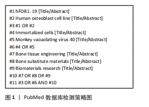
[1] KOONS GL, DIBA M, MIKOS AG. Materials design for bone-tissue engineering. Nat Rev Mater. 2020;5(8):584-603.
[2] 倪卓,杨莎,王应,等.骨替代材料的研究方法及进展[J].深圳大学学报(理工版),2015,32(4):331-342.
[3] 徐杨俊,赵建宁.体外培养成骨细胞的研究进展[J].中国骨伤,2010, 23(7):562-565.
[4] BRUN P. Culture of human primary bone cells and phenotype assessment. Methods Mol Biol. 2018;1727:413-421.
[5] 李楠,王和鸣.体外培养成骨细胞研究新进展[J].中国骨伤,2003, 16(3):63-65.
[6] HARRIS SA, ENGER RJ, RIGGS BL, et al. Development and characterization of a conditionally immortalized human fetal osteoblastic cell line. J Bone Miner Res. 1995;10(2):178-186.
[7] MAYO FOUNDATION FOR MEDICAL EDUCATION AND RESEARCH. Immortalized human fetal osteoblastic cells: US19960697906[P].1997-12-02.
[8] SUBRAMANIAM M, JALAL SM, RICKARD DJ, et al. Further characterization of human fetal osteoblastic hFOB 1.19 and hFOB/ER alpha cells: bone formation in vivo and karyotype analysis using multicolor fluorescent in situ hybridization. J Cell Biochem. 2002; 87(1):9-15.
[9] 程斌,程海燕.永生化骨髓间充质干细胞模型的构建[J].现代医院,2015,15(1):13-15.
[10] 张露文,戴鹏秀,张翊华,等.猴空泡病毒40大T抗原基因诱导人骨髓间充质干细胞的永生化[J].中国组织工程研究,2022,26(13): 1969-1973.
[11] 解慧琪,杨志明,屈艺.SV40与细胞永生化[J].中国修复重建外科杂志,2000,14(3):170-174.
[12] 陈文明,陈子兴,岑建农,等.人成骨细胞系hFOB1.19生物学特性的研究[J].中国实验血液学杂志,2008,16(2):339-344.
[13] OLIVEIRA PF, PINTO JP, SANTOS AR, et al. Evaluation of the growth and differentiation of human fetal osteoblasts (hFOB) cells on demineralized bone matrix (DBM). Organogenesis. 2021;17(3-4):136-149.
[14] TONG S, XU DP, LIU ZM, et al. Construction and in vitro characterization of three-dimensional silk fibroinchitosan scaffolds. Dent Mater J. 2015: 34(4):475-484.
[15] 郭乾臣.人骨肉瘤细胞株和成骨细胞株差异蛋白的比较蛋白质组学研究[D].广州:中山大学,2007.
[16] JO S, LEE JK, HAN J, et al. Identification and characterization of human bone-derived cells. Biochem Biophys Res Commun. 2018;495(1):1257-1263.
[17] MAROZIN S, SIMON-NOBBE B, IRAUSEK S, et al. Kinship of conditionally immortalized cells derived from fetal bone to human bone-derived mesenchymal stroma cells. Sci Rep. 2021;11(1):10933.
[18] STANLEY KT, VANDORT C, MOTYL C, et al. Immunocompetent properties of human osteoblasts: interactions with T lymphocytes. J Bone Miner Res. 2006;21(1):29-36.
[19] DOMINICI M, LE BLANC K, MUELLER I, et al. Minimal criteria for defining multipotent mesenchymal stromal cells. The International Society for Cellular Therapy position statement. Cytotherapy. 2006;8(4):315-317.
[20] YEN ML, CHIEN CC, CHIU IM, et al. Multilineage differentiation and characterization of the human fetal osteoblastic 1.19 cell line: a possible in vitro model of human mesenchymal progenitors. Stem Cells. 2007;25(1):125-131.
[21] 孟刚,李懿,辛榕,等.人永生化成骨细胞hFOB1.19恶性转化的研究[J].第三军医大学学报,2007,29(5):380-383.
[22] TONG S, XUE L, XU DP, et al. In vitro culture of hFOB1.19 osteoblast cells on TGF-beta1-SF-CS three-dimensional scaffolds. Mol Med Rep. 2016;13(1):181-187.
[23] MENG X, GONG K, SUN C, et al. Nonmineralized and mineralized silk fibroin/gelatin hybrid scaffolds: chacterization and cytocompatibility in vitro for bone-tissue engineering. J Craniofac Surg. 2020;31(2):416-419.
[24] FERREIRA CF, CARRIEL GOMES MC, FILHO JS, et al. Platelet-rich plasma influence on human osteoblasts growth. Clin Oral Implants Res. 2005; 16(4):456-460.
[25] TITORENCU I, ALBU MG, ANTONF, et al. Collagen-dexamethasone and collagen-d3scaffolds for bone tissue engineering. Mol Cryst Liq Cryst. 2012;555(1):208-217.
[26] CUI YC, QIU YS, WU Q, et al. Metabolic utilization of human osteoblast cell line hFOB 1.19 under normoxic and hypoxic conditions: a phenotypic microarray analysis. Exp Biol Med (Maywood). 2021; 246(10):1177-1183.
[27] LI B, HAN Y, LI M. Enhanced osteoblast differentiation and osseointegration of a bio-inspired HA nanorod patterned pore-sealed MgO bilayer coating on magnesium. J Mater Chem B. 2016;4(4):683-693.
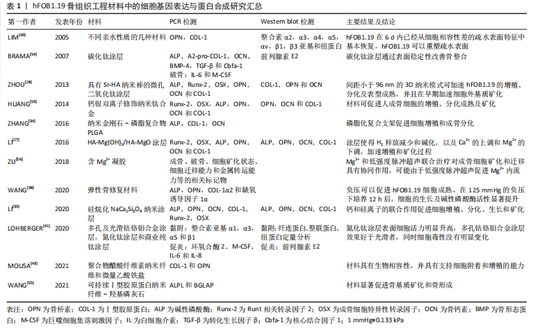
[28] ZHOU J, LI B, LU S, et al. Regulation of osteoblast proliferation and differentiation by interrod spacing of Sr-HA nanorods on microporous titania coatings. ACS Appl Mater Interfaces. 2013;5(11):5358-5365.
[29] LI B, HAN Y, QI K. Formation mechanism, degradation behavior, and cytocompatibility of a nanorod-shaped HA and pore-sealed MgO bilayer coating on magnesium. ACS Appl Mater Interfaces. 2014;6(20): 18258-18274.
[30] NOMOTO H, MAEHASHI H, SHIRAI M, et al. Bio-artificial bone formation model with a radial-flow bioreactor for implant therapy-comparison between two cell culture carriers: porous hydroxyapatite and beta-tricalcium phosphate beads. Hum Cell. 2019;32(1):1-11.
[31] MASAELI R, ZANDSALIMI K, TAYEBI L. Biomaterials evaluation: conceptual refinements and practical reforms. Ther Innov Regul Sci. 2019;53(1):120-127.
[32] HUAG HS, CHOU SH, DON TM, et al. Formation of microporous poly (hydroxybutyric acid) membranes for culture of osteoblast and fibroblast. Polym Adv Tech. 2009;20(12):1082-1090.
[33] GYORGY E, TORICELLI P, SOCOL G, et al. Biocompatible Mn2+-doped carbonated hydroxyapatite thin films grown by pulsed laser deposition. J Biomed Mater Res A. 2004;71(2):353-358.
[34] ZHANG F, SONG Q, HUANG X, et al. A novel high mechanical property PLGA composite matrix loaded with nanodiamond-phospholipid compound for bone tissue engineering. ACS Appl Mater Interfaces. 2016;8(2):1087-1097.
[35] LIU X, LIM JY, DONAHUE HJ, et al. Influence of substratum surface chemistry/energy and topography on the human fetal osteoblastic cell line hFOB 1.19:Phenotypic and genotypic responses observed in vitro. Biomaterials. 2007;28(31):4535-4350.
[36] TANG Y, ZHAO Y, WONG CS, et al. Apatite-coated three-dimensional fibrous scaffolds and their osteoblast response. J Biomed Mater Res A. 2013;101(3):674-683.
[37] PANDEY A, MIDHA S, SHARMA RK, et al. Antioxidant and antibacterial hydroxyapatite-based biocomposite for orthopedic applications. Mater Sci Eng C Mater Biol Appl. 2018;88:13-24.
[38] WANG R, THAYER P, GOLDSTEIN A, et al. Interaction of material stiffness and negative pressure to enhance differentiation of bone marrow-derived stem cells and osteoblast proliferation. J Tissue Eng Regen Med. 2020;14(2):295-305.
[39] LI K, XUE Y, ZHOU J, et al. Silanized NaCa2HSi3O9 nanorods with a reduced pH increase on Ti for improving osteogenesis and angiogenesis in vitro. J Mater Chem B. 2020;8(4):691-702.
[40] LIM JY, TAYLOR AF, LI ZY, et al. Integrin expression and osteopontin regulation in human fetal osteoblastic cells mediated by substratum surface characteristics. Tissue Eng. 2005;11(1-2):19-29.
[41] LOHBERGER B, STUENDL N, GLAENZER D, et al. CoCrMo surface modifications affect biocompatibility, adhesion, and inflammation in human osteoblasts. Sci Rep. 2020;10(1):1682.
[42] KUMAR A, HAN SS. Enhanced mechanical, biomineralization, and cellular response of nanocomposite hydrogels by bioactive glass and halloysite nanotubes for bone tissue regeneration. Mater Sci Eng C Mater Biol Appl. 2021;128:112236.
[43] MOUSA HM, HUSSEIN KH, SAYED MM, et al. Development and characterization of cellulose/iron acetate nanofibers for bone tissue engineering applications. Polymers (Basel). 2021;13(8):1339-1356.
[44] 刘晓峰,张郑瑶,田秀丽,等.纳米磷酸三钙/明胶/鹿茸多肽复合材料的制备及对人成骨细胞增殖、分化及矿化的影响[J].中国兽医学报,2013,33(6):898-904.
[45] BABUSKA V, PALAN J, KOLAJA DOBRA J, et al. Proliferation of osteoblasts on laser-modified nanostructured titanium surfaces. Materials (Basel). 2018;11(10):1827-1840.
[46] SMIGA-MATUSZOWICZ M, JANICKI B, JASZCZ K, et al. Novel bioactive polyester scaffolds prepared from unsaturated resins based on isosorbide and succinic acid. Mater Sci Eng C Mater Biol Appl. 2014;45:64-71.
[47] HAMDAN ALKHRAISAT M, MOSEKE C, BLANCO L, et al. Strontium modified biocements with zero order release kinetics. Biomaterials. 2008;29(35):4691-4697.
[48] TAI HY, CHOU SH, CHENG LP, et al. Asymmetric composite membranes from chitosan and tricalcium phosphate useful for guided bone regeneration. J Biomater Sci Polym Ed. 2012;23(9):1153-1170.
[49] ZO S, CHOI S, KIM H, et al. Synthesis and characterization of carboxymethyl chitosan scaffolds grafted with waterborne polyurethane. J Nanosci Nanotechnol. 2020;20(8):5014-5018.
[50] DONNADIO A, BINI M, CENTRACCHIO C, et al. Bioinspired reactive interfaces based on layered double hydroxides-zn rich hydroxyapatite with antibacterial activity. ACS Biomater Sci Eng. 2021;7(4):1361-1373.
[51] YUAN W, HE X, ZHOU X, et al. Hydroxyapatite nanoparticle-coated 3d-printed porous ti6al4v and cocrmo alloy scaffolds and their biocompatibility to human osteoblasts. J Nanosci Nanotechnol. 2018; 18(6):4360-4365.
[52] GU RL, LIU H, ZHU Y, et al. Is extracellular matrix (ECM) a promising scaffold biomaterial for bone repair? Histol Histopathol. 2021;36(12): 1219-1234.
[53] HUANG R, HAN Y, LU S. Enhanced osteoblast functions and bactericidal effect of Ca and Ag dual-ion implanted surface layers on nanograined titanium alloys. J Mater Chem B. 2014;2(28):4531-4543.
[54] BRAMA M, RHODES N, HUNT J, et al. Effect of titanium carbide coating on the osseointegration response in vitro and in vivo. Biomaterials. 2007;28(4):595-608.
[55] WANG JK, CIMENOGLUC, CHEAM NMJ, et al. Sustainable aquaculture side-streams derived hybrid biocomposite for bone tissue engineering. Mater Sci Eng C Mater Biol Appl. 2021;126:112104.
[56] ZU H, YI X, ZHAO D. Transcriptome sequencing analysis reveals the effect of combinative treatment with lowintensity pulsed ultrasound and magnesium ions on hFOB1.19 human osteoblast cells. Mol Med Rep. 2018;18(1):749-762.
[57] LU M, ZHAO XH. The growth proliferation, apoptotic prevention, and differentiation induction of the gelatin hydrolysates from three sources to human fetal osteoblasts (hFOB 1.19 Cells). Molecules. 2018;23(6):1287-1304.
[58] ZHANG Z, ZHANG X, ZHAO D, et al. TGFbeta1 promotes the osteoinduction of human osteoblasts via the PI3K/AKT/mTOR/S6K1 signalling pathway. Mol Med Rep. 2019;19(5):3505-3518.
[59] ZHANG X, ZU H, ZHAO D, et al. Ion channel functional protein kinase TRPM7 regulates Mg ions to promote the osteoinduction of human osteoblast via PI3K pathway: in vitro simulation of the bone-repairing effect of Mg-based alloy implant. Acta Biomater. 2017;63:369-382.
[60] KAZIMIERCZAK P, KOZIOL M, PRZEKORA A. The Chitosan/Agarose/NanoHA bone scaffold-induced M2 macrophage polarization and its effect on osteogenic differentiation in vitro. Int J Mol Sci. 2021;22(3): 1190-1204.
[61] KHALIFEHZADEH R, ARAMI H. DNA-templated strontium-doped calcium phosphate nanoparticles for gene delivery in bone cells. ACS Biomater Sci Eng. 2019;5(7):3201-3211.
[62] 孙中洋,李东韬,赵学武,等.成骨细胞体外培养模型的研究进展[J].中国骨质疏松杂志,2015,21(6):733-736,748.
[63] JIANG T, XU G, CHEN X, et al. Impact of hydrogel elasticity and adherence on osteosarcoma cells and osteoblasts. Adv Healthc Mater. 2019;8(9):e1801587.
[64] 孙亚东,张宏宇,华进联,等.胚胎干细胞体外分化为成骨细胞研究进展[J].中国修复重建外科杂志,2007,21(12):1381-1383.
[65] 殷晓雪,陈仲强,郭昭庆,等.转导人端粒逆生长细胞生长发育成骨生长的研究[J].中华医学杂志,2003,83(14):1251-1254.
[66] GHASEMI-MOBARAKEH L, KOLAHREEZ D, RAMAKRISHNA S, et al. Key terminology in biomaterials and biocompatibility. Curr Opin Biomed Eng. 2019;10:45-50.
[67] KOSKI C, VU AA, BOSE S. Effects of chitosan-loaded hydroxyapatite on osteoblasts and osteosarcoma for chemopreventative applications. Mater Sci Eng C Mater Biol Appl. 2020;115:111041.
[68] SARKAR N, BOSE S. Liposome-encapsulated curcumin-loaded 3d printed scaffold for bone tissue engineering. ACS Appl Mater Interfaces. 2019;11(19):17184-17192.
[69] LIU Y, QIAO Z, GAO J, et al. Hydroxyapatite-bovine serum albumin-paclitaxel nanoparticles for locoregional treatment of osteosarcoma. Adv Healthc Mater. 2021;10(2):e2000573.
[70] MAVER T, MASTNAK T, MIHELIC M, et al. Clindamycin-based 3d-printed and electrospun coatings for treatment of implant-related infections. Materials (Basel). 2021;14(6):1464-1481.
[71] VIDAL-GUTIERREZ X, PRADO-PRONE G, RODIL SE, et al. Bismuth subsalicylate incorporated in polycaprolactone-gelatin membranes by electrospinning to prevent bacterial colonization. Biomed Mater. 2021. doi: 10.1088/1748-605X/ac058d.
[72] HADRUP N, SHARMA A, LOESCHNER K. Toxicity of silver ions, metallic silver, and silver nanoparticle materials after in vivo dermal and mucosal surface exposure: a review. Regul Toxicol Pharmacol. 2018;98:257-267.
[73] RADHAKRISHNAN S, NAGARAJAN S, BELAID H, et al. Fabrication of 3D printed antimicrobial polycaprolactone scaffolds for tissue engineering applications. Mater Sci Eng C Mater Biol Appl. 2021;118:111525.
|