中国组织工程研究 ›› 2023, Vol. 27 ›› Issue (7): 1117-1125.doi: 10.12307/2023.044
• 生物材料综述 biomaterial review • 上一篇 下一篇
压电材料修复骨缺损的应用及设计思路
唐昊天,廖荣东,田 京
- 南方医科大学珠江医院关节骨病外科,广东省广州市 510280
-
收稿日期:2022-02-10接受日期:2022-03-02出版日期:2023-03-08发布日期:2022-07-20 -
通讯作者:田京,医学硕士,教授,博士生导师,现任珠江医院关节骨病外科教授、外科教研室主任、临床技能中心主任。 -
作者简介:唐昊天,男,1997年生,四川省绵阳市人,汉族,南方医科大学在读硕士,主要从事复杂骨病骨修复的组织工程治疗与临床研究。 -
基金资助:
Application and design of piezoelectric materials for bone defect repair
Tang Haotian, Liao Rongdong, Tian Jing
- Department of Orthopedics, Zhujiang Hospital, Southern Medical University, Guangzhou 510280, Guangdong Province, China
-
Received:2022-02-10Accepted:2022-03-02Online:2023-03-08Published:2022-07-20 -
Contact:Tian Jing, Master, Professor, Doctoral supervisor, Department of Orthopedics, Zhujiang Hospital, Southern Medical University, Guangzhou 510280, Guangdong Province, China -
About author:Tang Haotian, Master candidate, Department of Orthopedics, Zhujiang Hospital, Southern Medical University, Guangzhou 510280, Guangdong Province, China
摘要:
文题释义:
压电材料:某些在受到外力的作用而变形时,其内部会产生极化并使其表面产生电荷现象的晶体材料。骨作为一种天然的压电材料,通过将受到的应力转化为生物电信号,调节骨生长,结构重塑与修复。
细胞外基质:在多细胞有机体中,细胞周围由多种大分子组成的复杂网络。骨组织细胞外基质参与调节细胞黏附、增殖、分化,因此能够良好模拟细胞外基质的生物材料可促进骨修复。
背景:骨具有压电效应,可将其所受应力转化为骨表面的电信号,通过电信号调节骨的代谢与生长。具有生物电活性的压电材料作为骨植入物能在受力时表面生成电荷,恢复损伤骨组织表面电势从而达到促进骨愈合的目的。
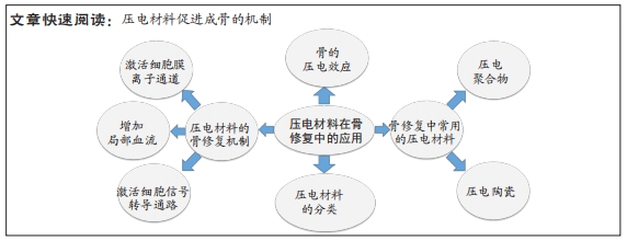
目的:文章从压电的角度介绍了骨的压电效应,说明压电材料促进骨缺损修复的可行性以及压电材料应用于骨组织工程的研究进展,并阐述压电材料促进成骨的机制,以期为骨缺损修复提供新的思路。
方法:以“Piezoelectric effect,Piezoelectric materials,Piezoelectric ceramic,Piezoelectric polymers,Osteogenesis,Bone tissue engineering,Scaffolds,Bone defect,energy harvester”为英文检索词,以“压电效应、压电材料、压电陶瓷、压电聚合物、成骨、骨组织工程、细胞支架、骨缺损、能量采集器”为中文检索词,在PubMed,Web of Science,ScienceDirect,中国知网和万方数据库中检索筛选出88篇文献进行归纳总结。
结果与结论:①压电材料具有因受力形变而产电的自发电特性,能模拟骨组织的细胞外基质的生物电微环境,可制备成生物支架,在不依赖于生长因子和药物的情况下给予骨组织电刺激,增强骨修复功能,在骨缺损的治疗中具有应用前景。②压电材料通过刺激细胞电压门控钙通道、α5β1整合素、促进局部血流增加等途径促进成骨。③目前尚未发现任何单一压电材料可满足组织工程的压电材料的特征,因此压电陶瓷与压电聚合物相结合,压电材料与生物活性材料相结合的压电复合材料应运而生,其中压电陶瓷-有机物复合材料,保留了压电陶瓷的压电性能优异和生物相容性良好优点的同时更易于加工,其生物相容性和促进骨细胞贴附、矿化等功能更强,是当前治疗骨缺损效果最好的压电材料;④虽然压电材料能模拟骨组织的生物电微环境来促进成骨,但由于对压电材料影响骨细胞的机制尚未明确,以及不同骨组织微环境各不相同,因此限制了压电材料在临床的应用,随着材料研发技术的进步和对压电材料作用于细胞的研究愈发深入,压电材料有望为骨缺损的修复提供新的思路。
https://orcid.org/0000-0003-1346-5037(唐昊天);https://orcid.org/0000-0001-9690-3850(田京)
中国组织工程研究杂志出版内容重点:生物材料;骨生物材料;口腔生物材料;纳米材料;缓释材料;材料相容性;组织工程
中图分类号:
引用本文
唐昊天, 廖荣东, 田 京. 压电材料修复骨缺损的应用及设计思路[J]. 中国组织工程研究, 2023, 27(7): 1117-1125.
Tang Haotian, Liao Rongdong, Tian Jing. Application and design of piezoelectric materials for bone defect repair[J]. Chinese Journal of Tissue Engineering Research, 2023, 27(7): 1117-1125.
使用本文
应用于骨组织工程的压电材料多为3类:压电聚合物,压电陶瓷,压电聚合物与压电陶瓷的复合物,材料详细分类见表1。
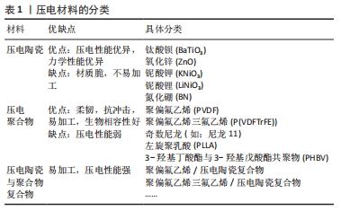
压电聚合物又分为天然压电聚合物和人工合成压电聚合物,天然压电聚合物包括纤维素、胶原和甲壳素,而人工合成压电聚合物包含聚偏氟乙烯以及它的共聚物聚偏氟乙烯三氟乙烯[poly(vinylidene fluorideand-trifluoroethylene)P(VDF-TrFE)]、奇数尼龙(如:尼龙11)、左旋聚乳酸(poly-L-lactic acid,PLLA)、3-羟基丁酸酯与 3-羟基戊酸酯共聚物(poly-3-hydroxybutyrate-3-hydroxy valerate,PHBV)等。压电聚合物具有良好的柔韧性、易加工性、抗冲击性和生物相容性,在组织工程领域得到广泛的应用。压电陶瓷是一种多晶体,如钛酸钡(barium titanate,BaTiO3)、铌酸钾钠(potassium sodium niobate,KNN)和铌酸锂钠钾(lithium sodium potassium niobate,LKNN)、氧化锌(Zinc oxide,ZnO),氮化硼纳米管(boron nitride nanotubes,BNNT)等材料,这一类压电材料相较于压电聚合物具有更强的压电性能。压电复合材料则是将压电陶瓷与聚合物通过复合工艺而制成的新型材料。压电陶瓷压电性能优异,但其脆性和不易加工性限制了其单独在骨组织工程中的应用[17]。压电聚合物具有良好的柔韧性、抗冲击性、易加工性和生物相容性,但其压电性能较弱,难以达到刺激细胞的阈值。开发聚合物-陶瓷复合材料有望结合它们的优点,克服各自的限制,研发出性能更加优异的压电材料[18]。
2.2 骨的压电效应 骨组织是一种具有再生能力的组织,骨修复也是一个复杂的过程,依赖于机械、化学、生物电等生物环境[19]。人体的骨组织是一种天然的压电材料。日本科学家于1954年首次报道了骨具有压电性[20]。人体骨骼由细胞和细胞外基质组成,其中细胞包含了成骨细胞、破骨细胞、骨细胞和骨髓细胞(包括造血干细胞),而细胞外基质由65%的矿物质(羟基磷灰石为主)与35%胶原组成,Ⅰ型胶原约占胶原的90%,具有三重螺旋结构,为骨骼提供主要的拉伸强度。而无机物则以羟基磷灰石钙的形式为骨骼提供主要的抗压强度。最早的研究认为,骨的压电效应主要来源于胶原分子。胶原是一种纤维性蛋白,FUKADA[21]的研究提出剪切力使胶原纤维相互错动进而发生极化,使骨骼体现出压电性能。骨的压电效应使骨胶原在受力发生形变时,其压缩侧呈负电位,张力侧呈正电位。天然骨骼具有固有的Zeta电位为-5 mV,而由于骨胶原的压缩而产生的负电荷增加了自然活骨中的总Zeta电位和流动电位[22]。除了胶原纤维,骨的压电性能和羟基磷灰石也有关。LANG等[23]通过对应压电力显微镜观察发现了羟基磷灰石具有压电性,该实验结果表明,羟基磷灰石对骨的压电性具有一定贡献。根据沃尔夫定律,骨可以对机械负载做出反应,在应力较大的区域骨的生长和重塑能力增强[24],其原因在于机械应力增加了骨压电电位的负电荷 [25]。在一定范围内,骨所受压力负荷越高,骨表面负电位越高,促进成骨细胞增殖分化的效果越强,成骨作用越强,这也很好地解释了为什么长期卧床的患者和太空旅行的宇航员往往骨密度低,易患骨质疏松。有研究表明,骨组织在受到应力时,带负电位的一面成骨细胞增殖分化能力加快,该面呈骨修复状态,带正电位一面的破骨细胞活性增强,该面呈骨吸收状态[26]。因此,骨的压电效应在骨修复中起重要作用。
2.3 骨修复中常用的压电材料 压电材料是电活性材料,具有成为新型骨替代材料的巨大潜力。压电材料将自身受到的压力转化为合适的电刺激,具有成为骨替代材料的潜力。骨修复研究中的压电材料除了应具备优异的电学性能以外,还应有良好的生物相容性和机械性。
2.3.1 压电聚合物 压电聚合物质地与无机晶体相比,其柔韧性好,易于加工。当前,应用于组织工程学的压电聚合物满足以下条件:①存在永久的分子偶极子;②拥有校准或对齐分子偶极子的能力;③具有维持偶极子对齐状态的能力;④聚合物在受到机械应力时抗牵张的能力[27]。压电聚合物大多生物相容性良好,但机械强度常不足,难以作为骨植入物承受生理负荷。并且聚合物压电性能不如压电陶瓷,因此单独采用压电聚合物可能难以达到促进骨修复的电刺激阈值。目前多推荐聚合物与无机陶瓷结合形成压电复合材料,一方面增强自身的机械强度,另一方面复合材料本身会使聚合物生物相容性增加,这可能是材料压电性能增强从而促进了细胞的贴附、增殖与分化功能。
组织工程研究中常见的压电聚合物如下:
聚偏氟乙烯:是当前研究最广泛的压电共聚物,其压电常数为34 pC/N。由于聚偏氟乙烯柔韧性高,无细胞毒性,因此广泛应用于生物医学领域并被研制出多种产品,包括了组织工程支架和可植入的自充电设备。当前已有研究证实压电聚合物聚偏氟乙烯基底与小鼠颅顶前成骨细胞 (MC3T3-E1)、人间充质干细胞、人脂肪干细胞和山羊骨髓干细胞的生物相容性[28-31]。极化的聚偏氟乙烯压电薄膜能产生足够的电势,诱导前成骨细胞的增殖分化。MARINO等[32]报告,当极化与未极化的聚偏氟乙烯聚合物基质分别植入大鼠胫骨骨间膜6周时,极化的聚偏氟乙烯成骨性能显著高于未极化聚偏氟乙烯。有学者报道聚偏氟乙烯共聚物和钛酸钡压电复合物相结合时能显著提高聚偏氟乙烯的压电性能,同时也能解决钛酸钡质脆、难加工的问题[33]。RIBEIRO等[34]制作的表面极化的β-聚偏氟乙烯薄膜同普通聚偏氟乙烯薄膜相比,能够提高人骨髓间充质干细胞碱性磷酸酶碱性磷酸酶的活性,同时薄膜在受到动态的机械振动时,其促成骨的效果会进一步增强。REIS等[35]报道了一种基于聚偏氟乙烯的新型压电装置能够利用逆压电效应有效地刺激骨-种植体界面处的骨生长。
当前普遍认为聚偏氟乙烯是具有良好生物相容性的热塑性聚合物,具有极高的耐腐蚀性,虽然其在极端碱性环境会降解,但在正常的生物环境下是不可降解的材料,这也因此限制了其在组织工程学的广泛应用[36]。
聚偏氟乙烯-三氟乙烯:是偏氟乙烯和三氟乙烯的共聚物,是当前压电常数最高的压电聚合物(38 pC/N)。偏氟乙烯和三氟乙烯共聚物具有良好的细胞生物相容性并对细胞的贴附和分化有促进作用[37],可促进骨、皮肤、软骨和肌腱等多种组织的再生。DAMARAJU等[38]通过静电纺丝技术制造了偏氟乙烯和三氟乙烯共聚物的3D支架,并证明了与非压电材料相比,压电性和机械应力的联合作用更能刺激人骨髓间充质干细胞的增殖、分化、细胞外基质矿化和基因表达。DAI等[39]制备的聚偏氟乙烯-三氟乙烯-钛酸钡压电复合膜可模拟骨组织生物电微环境,通过炎症介导的机制,促进糖尿病大鼠的骨再生。聚合物混合物在骨和软骨组织工程的应用的正变得越来越重要,而聚偏氟乙烯和聚偏氟乙烯-三氟乙烯与淀粉等天然聚合物结合,其形成的聚偏氟乙烯-淀粉和聚偏氟乙烯和三氟乙烯-淀粉植入物异物反应少,弹性模量与松质骨相当,可作为骨修复支架,具有应用于骨组织工程的潜力[40]。LOPES等[41]比较了聚偏氟乙烯和三氟乙烯-10%钛酸钡压电复合材料和聚四氟乙烯分别植入大鼠颅骨缺损4周和8周后的新骨生成量,与聚四氟乙烯相比,偏氟乙烯和三氟乙烯共聚物-10%钛酸钡压电复合材料的表面成骨显著增加,这清楚地表明了压电基底的促进骨生长的效果,这可能是因为聚偏氟乙烯和三氟乙烯-10%钛酸钡压电复合材料通过其压电性、亲水性从而增强蛋白结合能力,促进骨生成。而聚偏氟乙烯和三氟乙烯与氮化硼纳米管形成的偏氟乙烯和三氟乙烯共聚物- 氮化硼纳米管压电复合材料相比偏氟乙烯和三氟乙烯共聚物其压电性能提高近2倍,因此偏氟乙烯和三氟乙烯共聚物- 氮化硼纳米管压电复合材料的骨生成量会增加[42]。
聚-3-羟基丁酸-3-羟基戊酸酯:是聚羟基脂肪酸家族的一员,于1986年问世,由于其良好的生物相容性、可生物降解性和热塑性,在生物医学领域的地位逐步提高[43]。聚-3-羟基丁酸-3-羟基戊酸酯,其通过酶降解机制水解并释放二氧化碳,与其他生物聚合物相比,其生物相容性良好,植入动物体内引起的炎症反应发生率更低,并且其压电常数(1.3 pC/N)与人骨相似,以上特性使其具有模拟自然骨及作为骨植入物的潜力[44]。
聚3-羟基丁酸-3-羟基戊酸酯由于生物活性以及亲水性差,因此需要通过研发与其他材料相结合的复合压电材料来克服聚(3-羟基丁酸-3-羟基戊酸酯)的缺点。可生物降解的聚-3-羟基丁酸-3-羟基戊酸酯-羟基磷灰石复合材料已被证明具有促进成骨的功能,因此 GORODZHA等[45]成功制备了由聚(3-羟基丁酸-3-羟基戊酸酯)与含硅酸盐的羟基磷灰石组成的复合材料骨支架,该支架促进了人骨髓间充质干细胞的增殖、贴附与分化功能。有研究制备的聚-3-羟基丁酸-3-羟基戊酸酯-壳聚糖-羟基磷灰石支架相较于普通聚-3-羟基丁酸-3-羟基戊酸酯支架其生物相容性显著提高,并有效促进成骨细胞贴附、增殖与分化[46]。
总之,聚羟基脂肪酸家族可与无机材料、聚合物材料和生物活性材料相结合形成复合材料从而达到改善自身生物活性与亲水性差的问题[47]。当前对于聚(3-羟基丁酸-3-羟基戊酸酯)混合材料研发有助于不断完善单一材料自身的缺陷,以实现材料之间促进成骨效应的协同作用。通过在未来混合材料制作的统一标准出台,聚(3-羟基丁酸-3-羟基戊酸酯)有望制成可生物降解的复合支架,用来治疗骨缺损。
聚酰胺:又称为尼龙,作为最早工业化的高分子材料已有50多年的历史,由于密度小、耐磨、耐油、易加工、特别好的力学性能而广泛应用于机械、交通、电气、化工、纺织及日用品方面。而奇术尼龙(尼龙-5,尼龙-7),其每个单体单元上含有偶数个亚甲基和一个酰胺基,因为自身的一个酰胺键可以产生净偶极矩(3.7 D)从而具有压电性。尼龙的压电常数(d31)在25 ℃时为3 pC/N,在107 ℃时为14 pC/N[48]。WANG等[49]报道,聚酰胺-羟基磷灰石复合材料在植入12周后可促进成骨。聚酰胺可作为聚合物基质用于软骨修复或再生,但由于其细胞相容性一般,所以需要适当的修饰以促进软骨细胞的细胞附着和增殖。由于聚酰胺缺乏降解模式使得其在组织工程中的应用受到限制。
左旋聚乳酸:半晶态左旋聚乳酸可以在未极化的状态下产生压电性,其原因在于左旋聚乳酸受到机械应力时,其分子中的中C=O键的会发生机械移位,导致净偶极矩和电荷的出现[50]。IKADA等[51]使用左旋聚乳酸作为骨替代物,并证明了左旋聚乳酸的极化压电效应能促进骨的生成。薄膜和杆状的压电左旋聚乳酸基板已被证实具有骨组织潜在替代物的适用性[52]。含有磷灰石和胶原的左旋聚乳酸支架在细胞外培养基中能促进人成骨肉瘤细胞(SaOS-2细胞)和人胎儿成骨细胞的代谢[53]。KO等[54]通过静电纺丝技术制备了左旋聚乳酸-脱矿骨粉纳米纤维复合支架,并在大鼠模型中观察到左旋聚乳酸-脱矿骨粉纳米纤维复合支架能有效促进人骨髓间充质干细胞的成骨分化,有效修复颅骨缺损。左旋聚乳酸由于具有可生物降解的特点,因此材料加工会受到限制,而左旋聚乳酸与淀粉形成淀粉共混物能增加左旋聚乳酸的延展性和压缩模量从而改善左旋聚乳酸加工性能的问题[55]。左旋聚乳酸具有无细胞毒性、可生物降解、材质适合骨科手术植入等优势,有望制备成可降解螺钉、固定钉与缝合锚钉从而避免植入物需行二次手术取出的问题[56]。
当前左旋聚乳酸生物相容性良好,可生物降解,植入体内不良反应少,机械强度高,但因其具有疏水性不利于细胞贴附,降解产生酸性产物能引起机体局部炎症反应,以及降解时间过长,往往超过骨愈合时间,因此左旋聚乳酸需要与其他材料如纳米陶瓷材料、碱金属材料相结合,改善自身的疏水性,减弱降解产生不良反应,并将生物降解时间控制在有利于骨与血管长入支架的合理区间,这样才有助于将左旋聚乳酸应用于骨缺损修复。
2.3.2 压电陶瓷 目前常见的压电陶瓷包括了锆钛酸铅(lead zirconate titanate,PZT)、钛酸钡、氧化锌(ZnO)、铌酸钾钠(KNN)、铌酸锂钠钾(lithium sodium potassium niobate,LNPN)和氮化硼纳米管(boron nitride nanotubes),其相较于压电聚合物的特点是压电常数高。评估压电陶瓷能否应用于骨组织工程的重要指标就是材料的细胞毒性。总体而言,含铅的压电陶瓷如锆钛酸铅,具有细胞毒性,因此在组织工程的材料研发与应用中较为少见。其他无铅的压电陶瓷的细胞毒性与材料的含量相关,相较于含铅压电陶瓷,在组织工程的研究中更受关注。总体而言,钛酸钡中所含钛与钡具有细胞毒性,氧化锌分解产物活性氧具有细胞毒性,而氮化硼、铌酸钾钠等材料所含元素无细胞毒性。但当前压电陶瓷往往与其他材料结合形成压电复合材料,通过改良与修饰可以克服陶瓷自身的不足[57-79]。
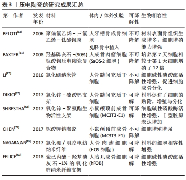
目前压电聚合物研究成果见表2,压电陶瓷研究成果见表3。

钛酸钡(BaTiO3):钛酸钡由于生物相容性良好、压电性能优异同时具有满足骨骼生理负荷的机械强度,成为了目前研究最多的压电材料[57]。钛酸钡的结构在居里温度(120 ℃)以下其形态会发生从非极性的对称立方到极性的不对称立方的转变,这使得钛酸钡会产生自发的电极化[58]。钛酸钡是最早作为骨植入物治疗骨缺损的压电材料,有研究者于1980年将钛酸钡植入狗的股骨内,最后观察到了骨组织在材料表面矿化结果良好,未见植入物引起的炎症与排斥反应,证明了钛酸钡良好的生物相容性。随着对钛酸钡的性能不断研究,发现钛酸钡压电性能优异,即使少量应用也可满足骨生长所需电微环境,因此目前钛酸钡常常少量地作为填充物或表面涂层应用于压电材料中,例如羟基磷灰石-钛酸钡,聚偏氟乙烯-钛酸钡等。
目前已有研究支持了具有生物相容性的羟基磷灰石-钛酸钡压电陶瓷在骨种植支架开发的巨大潜能。BAXTER等[60]证明人成骨肉瘤细胞(SaOS-2细胞)在羟基磷灰石-(90%)钛酸钡压电陶瓷复合物(d33=57.8 pC/N)上培养,培养第7天细胞相较于第1天细胞增殖了12倍。羟基磷灰石-钛酸钡复合物的压电性能与钛酸钡的含量有关,钛酸钡含量越高,材料压电性能越强,当钛酸钡含量低于70%,复合物便失去压电性[61]。DUBEY等[62]在小鼠膝关节内注射羟基磷灰石-40%钛酸钡颗粒。注射7 d后,小鼠的活动和体质量正常,组织病理学分析表明,注射颗粒既没有转移到任何主要器官,如心脏、肾脏、肝脏、肺等,也没有对这些器官造成任何不良影响。而聚集在膝关节区域的注射颗粒并未造成任何炎症与异物反应,这表明羟基磷灰石-钛酸钡生物相容性良好。钛酸钡诱导的压电聚合物能显著提高聚合物的生物相容性并且能有效促进体内与体外的成骨效应。BELOTI等[63]观察到压电聚合物薄膜[聚偏氟乙烯-三氟乙烯-钛酸钡]促进人成骨细胞增殖分化的能力强于非压电聚合物聚四氟乙烯,这是由于压电聚合物薄膜[聚偏氟乙烯-三氟乙烯-钛酸钡]的亲水性与蛋白结合能力更强。
从上述研究可发现,钛酸钡压电陶瓷能产生压电电位,刺激羟基磷灰石的生成,增强细胞功能,在动物模型中展现出良好的生物相容性。但钛酸钡单独作为骨植入物具有局限性,具体表现在钡离子与钛离子具有细胞毒性,且材料自身的生物惰性阻碍了细胞贴附以及不可降解,导致了钛酸钡不可长期植入体内。因此,研究者应该利用钛酸钡自身压电优异的性能,高机械强度的优势,与其他材料结合,用来改善其他骨植入材料的性能,例如与羟基磷灰石相结合以提高材料的机械强度,与钛合金相结合以提高钛合金的生物相容性和压电性能,与聚合物相结合提高通过提高材料压电效应从而提高材料生物相容性。随着对钛酸钡的研究不断深入,钛酸钡有成为新一代骨植入物的潜能。
氧化锌(ZnO):锌通过调节转录因子、金属蛋白酶和聚合酶等不同酶的活性调节细胞代谢,是细胞增殖和分化中的重要元素。氧化锌生物相容性好,抗菌性能好,是良好的组织工程材料。纳米氧化锌具有压电效应,在受到细胞自身固有机械应力时能产生局部电位,显著改善人SaOS-2成骨样细胞和巨噬细胞的代谢活性[64]。LAURENTI等[65]提出,对于特定的固定纳米尺寸,高浓度(60 μg/mL)的氧化锌纳米颗粒与低浓度(30 μg/mL)相比更容易降低骨髓间充质干细胞的生物活性,这表明氧化锌具有浓度依赖性的细胞毒性。除了浓度,氧化锌的细胞毒性还取决于颗粒直径和孔隙密度。由于纳米氧化锌会生成活性氧,随着颗粒直径的减小,活性氧生成增加,细胞毒性增强。随着孔隙密度的增加,纳米氧化锌供蛋白反应以及细胞贴附的表面积增加,成纤维细胞的功能也随之提升[66]。大量文献研究表明,在生物材料中加入适量的氧化锌可以促进材料与成纤维细胞、成骨细胞、干细胞等的生物相容性[65-67]。例如,DIKICI等[67]报告,随着硫酸钙支架中氧化锌含量增加至0.5%,材料表面的人骨髓间充质干细胞密度也随之升高,该现象可归因于压电氧化锌能增强支架的亲水性,从而利于细胞的贴附。适量的压电氧化锌也能提高聚合物支架(如聚己内酯和聚氨酯)的成骨反应。SHRESTHA等[68]通过引入体积分数0.2%氧化锌纳米颗粒,在聚氨酯聚合物支架上观察到前成骨细胞MC3T3-E1的刺激成骨增殖和分化。据报道,与不含氧化锌的支架相比,通过在聚己内酯-羟基磷灰石中加入体积分数1%的氧化锌纳米纤维支架能增强人胎儿成骨细胞活性 [69]。纳米氧化锌改善生物材料相容性的原因可能与锌离子的可持续释放相关,而释放的锌离子可调节各种细胞代谢活动,如蛋白质合成和mRNA表达等[70]。
氧化锌生物相容性好,展现出良好的压电性,自身生成的活性氧一方面具有抗菌效果,另一方面会产生剂量依赖的细胞毒性,这是不可忽视的。因此一方面,研究者在用氧化锌进行临床试验前,应该在动物模型中寻找氧化锌的最适浓度,另一方面应通过对氧化锌材料进行改进和修饰,从而使氧化锌能够作用于骨组织工程。
铌酸钾钠(KNN)和铌酸锂钠钾(LKNN):作为无铅的压电材料,其中包含的金属铌为人体必需微量元素,具有无细胞毒性、生物相容性好的特点,有学者利用铌酸钾钠的压电性能将其制为纳米药物输送设备,从而达到促进骨与软骨修复与再生的目的[71]。CHEN等[72]报道了表面极化的铌酸钾钠陶瓷材料相较于非极化陶瓷材料均能促进细胞蛋白吸收,无论是正电极化还是负电极化,但该实验中小鼠颅顶前成骨细胞(MC3T3-E1) 在负电位极化表面细胞密度显著高于正电位极化与非极化表面。铌酸锂钠钾具有化学稳定性与亲水的特点,这意味着其具有应用于骨科的价值[73]。当前利用铌酸钾钠诱导压电性的羟基磷灰石-铌酸钾钠梯度功能材料研发受到关注。羟基磷灰石-铌酸钾钠是将铌酸钾钠材料置于羟基磷灰石层之间,这样可以在不影响羟基磷灰石自身生物相容性的同时将羟基磷灰石的极化率提高3倍以上,而铌酸锂钠钾置于羟基磷灰石层之间可以使羟基磷灰石极化率提高6倍[73]。
正如上述研究所述,材料的生物相容性与自身的细胞毒性和表面极化有很大关系,而羟基磷灰石极化率的增加可能会促进细胞的增殖和分化。铌酸钾钠在受到生理负荷的情况下产生表面电荷从而达到极化的效果,能促进细胞的贴附与增殖,同时铌酸钾钠与铌酸锂钾钠自身无细胞毒性,两者相互作用使材料具有良好的生物相容性,有助于骨的再生。
氮化硼纳米管(BNNTs):硼,在分子水平上通过调节骨形态发生蛋白、骨钙素、RunX2(转录因子)的水平,在调节小鼠颅顶前成骨细胞(MC3T3-E1)和人骨髓间充质干细胞的成骨功能中发挥重要作用[74-75]。例如,NAKHMANSON等[76]研究了氮化硼纳米管(氮化硼纳米管)的压电性和自发极化的特点。氮化硼纳米管通过在培养基中释放的微量硼和氮化硼纳米管纤维对人骨髓间充质干细胞施加的使细胞受到拉伸,激活肌动蛋白丝的应力,促进人骨髓间充质干细胞向成骨细胞分化[77]。促进氮化硼纳米管成骨的另一个因素是其对细胞蛋白的亲和力强[78]。通过在明胶聚合物上添加质量分数1%–5%的氮化硼制备的静电纺纳米纤维,能使聚合物具备良好的促生物矿化作用和与人骨肉瘤细胞的相容性[79]。
随着纳米技术的发展,纳米结构的陶瓷材料开始出现。当前的压电聚合物大多生物相容性优于压电陶瓷且易于加工处理,但由于其自身机械强度与压电性能不足导致了其难以单独作为骨植入物。氮化硼纳米管仅需少量的加入便可提升压电聚合物的机械强度和压电性能,从而使复合材料能够更加符合骨植入物的力学要求。总的来说,上述研究清楚地表明了氮化硼在骨组织工程应用中的潜力。
2.4 压电材料优缺点总结 单一的压电材料在应用于骨修复缺损时都存在自身的限制与不足。应用于骨缺损修复的骨植入物需要在力学性质上满足承重要求,在细胞生物学上需要具备良好的生物活性,以利于细胞的贴附生长与分化。当前压电复合材料是组织工程的研究热点,其优势在于将不同压电材料相结合从而弥补单一材料的缺陷,整合不同材料的优点,使得制备理想的骨修复材料成为可能。为了实现此目的,一方面需要利用先进的技术手段改进材料的理化性质,通过多次实验以使材料压电电位达到可增强细胞生物相容性的临界值;另一方面便是了解不同压电材料的优缺点、不良反应、生物相容性,方可在材料的研发阶段达到优势互补的效果,从而制备出促进骨缺损修复的新型骨植入物。压电材料的优缺点总结见表4。

2.5.3 增加局部血流 电刺激通可促进血管扩张,增强血管通透性并增加局部组织血流。电刺激可以激发骨折坏死处邻近的血管内皮细胞形成新生血管并长入缺血的断端,为损伤组织提供血供。研究证实直流电刺激促进血管内皮细胞的生成新血管的同时,可通过血管内皮生长因子介导的反馈通路选择性调节血管生长因子和细胞因子在血管生成中的重要作用[88]。

| [1] CHEN R, WANG J, LIU C. Biomaterials act as enhancers of growth factors in bone regeneration. Adv. Funct. Mater. 2016;26(48):8810-8823. [2] 熊莹,许燕,周建平,等.组织工程研究中的电活性生物材料[J].中国组织工程研究,2019,23(34):5223-5330. [3] VASQUEZ-SANCHO F, ABDOLLAHI A, DAMJANOVIC D, et al. Flexoelectricity in bones. Adv Mater. 2018;30(9):1705316.1-1705316.5. [4] KIM D, HAN SA, KIM JH, et al. Biomolecular piezoelectric materials:from amino acids to living tissues. Adv Mater. 2020;32(14):1906989.1-1906989.16. [5] CHERNOZEM RV, SURMENEVA MA, ABALYMOV AA, et al. Piezoelectric hybrid scaffolds mineralized with calcium carbonate for tissue engineering: analysis of local enzyme and small-molecule drug delivery, cell response and antibacterial performance. Mater Sci Eng C Mater Biol Appl. 2021;122:111909.1-111909.10. [6] TANDON B, BLAKER JJ, CARTMELL SH, et al. Piezoelectric materials as stimulatory biomedical materials and scaffolds for bone repair. Acta Biomater. 2018;73(4):1-20. [7] RIBEIRO C, CORREIA DM, RODRIGUES I, et al. In-vivo demonstration of the suitability of piezoelectric stimuli for bone reparation. Materials Letters. 2017; 209(7):118-121. [8] 刘国峰.自供电刺激骨支架的激光增材制造及其性能研究[D].南昌:江西理工大学,2021. [9] BUI TT, SHIN MK, JEE SY, et al. Ferroelectric PVDF nanofiber membrane for high-efficiency PM0.3 air filtration with low air flow resistance. Colloids Surf A Physicochem Eng Asp. 2022;640:128418. [10] RAHMAN T, MARTIN NP, JENKINS JK, et al. Nb2O5, LiNbO3, and (Na, K)NbO3 Thin Films from High-Concentration Aqueous Nb-Polyoxometalates. Inorg Chem. 2022;61(8):3586-3597. [11] WEI H, GENG W, BI K, et al. High-performance piezoelectric-type MEMS vibration sensor based on LiNbO3 single-crystal cantilever beams. Micromachines (Basel). 2022;13(2):329. [12] BASSETT CA, PAWLUK RJ, BECKER RO. Effects of electric currents on bone in vivo. Nature. 1964;204(4959):652-654. [13] CARTER A, POPOWSKI K, CHENG K, et al. Enhancement of bone regeneration through the converse piezoelectric effect, a novel approach for applying mechanical stimulation. Bioelectricity. 2021;3(4):255-271. [14] PARK JB, KELLY BJ, KENNER GH, et al. Piezoelectric ceramic implants:in vivo results. J Biomed Mater Res. 1981;15(1):103-110. [15] CIOFANI G, RICOTTI L, CANALE C, et al. Effects of barium titanate nanoparticles on proliferation and differentiation of rat mesenchymal stem cells. Colloids Surf B Biointerfaces. 2013;102:312-320. [16] LI J, YANG F, LONG Y, et al. Bulk ferroelectric metamaterial with enhanced piezoelectric and biomimetic mechanical properties from additive manufacturing. ACS Nano. 2021;15(9):14903-14914. [17] MAIA FR, BASTOS AR, OLIVEIRA JM. Recent approaches towards bone tissue engineering. Bone. 2021;154:116256. [18] KAO FC, CHIU PY, TSAI TT, et al. The application of nanogenerators and piezoelectricity in osteogenesis. Sci Technol Adv Mater. 2019;20(1):1103-1117. [19] BRADY MA, WALDMAN SD, ETHIER CR. The application of multiple biophysical cues to engineer functional neocartilage for treatment of osteoarthritis. Part I: cellular response. Tissue Eng Part B Rev. 2015;21(1):1-19. [20] JARKOV L, ALLAN SJ, BOWEN C, et al. Piezoelectric materials and systems for tissue engineering and implantable energy harvesting devices for biomedical applications. Int Mater Rev. 2021. doi:10.1080/09506608.2021.1988194. [21] FUKADA E. Piezoelectric properties of biological polymers. Q Rev Biophys. 1983; 16(1):59-87. [22] AHN AC, GRODZINSKY AJ. Relevance of collagen piezoelectricity to “Wolff’s Law”: a critical review. Med Eng Phys. 2009;31(7):733-741. [23] LANG S, TOFAIL S, KHOLKIN A, et al. Ferroelectric polarization in nanocrystalline hydroxyapatite thin films on silicon. Sci Rep. 2013;3(1):1-6. [24] WANG Q, YANG J, ZHANG W, et al. Manufacture and cytotoxicity of a lead-free piezoelectric ceramic as a bone substitutc consolidation of porous lithium sodium potassium niobate by cold isostatic pressing. Int J Oral Sci. 2009;5(2):99-104. [25] HASTINGS GW, MAHMUD FA, Electrical effects in bone. J Biomed Eng. 1988;10(6): 515-521. [26] ZHENG T, HUANG Y, ZHANG X, et al. Mimicking the electrophysiological microenvironment of bone tissue using electroactive materials to promote its regeneration. J Mater Chem B. 2020;8(45):10221-10256. [27] GUO W, TAN C, SHI K, et al. Wireless piezoelectric devices based on electrospun PVDF/BaTiO3 NW nanocomposite fibers for human motion monitoring. Nanoscale. 2018;10(37):17751-17760. [28] RIBEIRO C, SENCADAS V, CORREIA DM, et al. Piezoelectric polymers as biomaterials for tissue engineering applications. Colloids Surf B Biointerfaces. 2015;136(8):46-55. [29] DAMARAJU SM, WU S, JAFFE M, et al. Structural changes in PVDF fibers due to electrospinning and its effect on biological function. Biomed Mater. 2013; 8(4):045007. [30] PARSSINEN J, HAMMAREN H, RAHIKAINEN R, et al. Enhancement of adhesion and promotion of osteogenic differentiation of human adipose stem cells by poled electroactive poly (vinylidene fluoride). J Biomed Mater Res. 2015;103(3):919-928. [31] RODRIGUES MT, GOMES ME, MANO JF, et al . β- membranes induce cellular proliferation and differentiation in static and dynamic conditions. Mater Sci Forum. 2008;587-588:72-76. [32] MARINO A, ROSSON J, GONZALEZ E, et al. Quasi static charge interactions in bone. J Electrost. 1988;21:347-360. [33] ZHANG CG, LIU WW, CAO C, et al. Modulating surface potential by controlling the β phase content in poly (vinylidene fluoridetrifluoroethylene) membranes enhances bone regeneration. Adv Healthc Mater. 2018;7(11):e1701466. [34] RIBEIRO C, PÄRSSINEN J, SENCADAS V, et al. Dynamic piezoelectric stimulation enhances osteogenic differentiation of human adipose stem cells. J Biomed Mater Res A. 2015;103(6):2172-2175. [35] REIS J, FRIAS C, CANTO E CASTRO C, et al. A new piezoelectric actuator induces bone formation in vivo:a preliminary study. Biomed Res Int. 2012;2012:613403. [36] NEUSS S, APEL C, BUTTLER P, et al. Assessment of stem cell/biomaterial combinations for stem cell-based tissue engineering. J Biomater. 2008;29(3):302-313. [37] WEBER N, LEE YS, SHANMUGASUNDARAM S, et al. Characterization and in vitro cytocompatibility of piezoelectric electrospun scaffolds. Acta Biomater. 2010; 6(9):3550-3556. [38] DAMARAJU SM, SHEN Y, ELELE E, et al. Three-dimensional piezoelectric fibrous scaffolds selectively promote mesenchymal stem cell differentiation. J Biomater. 2017;149(9):51-62. [39] DAI XH, HENG BC, BAI YY, et al. Restoration of electrical microenvironment enhances bone regeneration under diabetic conditions by modulating macrophage polarization. Bioact Mater. 2020;6(7):2029-2038. [40] PEREIRA JD, CAMARGO RC, FILHO CJ, et al. Biomaterials from blends of fluoropolymers and corn starchimplant and structural aspects. Mater Sci Eng. 2014;36(12):226-236. [41] LOPES HB, SANTOS TDS, OLIVEIRA FSD, et al. Poly (vinylidene-trifluoroethylene)/barium titanate composite for in vivo support of bone formation. J Biomater Appl. 2014;29(1):104-112. [42] GENCHI GG, SINIBALDI E, CESERACCIU L, et al. Ultrasound-activated piezoelectric P (VDFTrFE)/boron nitride nanotube composite films promote differentiation of human SaOS-2 osteoblast-like cells. Nanomed Nanotechnol Biol Med. 2018;14(7): 2421-2432. [43] PRYADKO A, SURMENEVA MA, SURMENEV RA, et al. Review of hybrid materials based on polyhydroxyalkanoates for tissue engineering applications. Polymers (Basel). 2021;13(11):1738. [44] CHERNOZEM RV, SURMENEVA MA, SHKARINA SN, et al. Piezoelectric 3-D fibrous poly (3-hydroxybutyrate)-based scaffolds ultrasound-mineralized with calcium carbonate for bone tissue engineering: inorganic phase formation, Osteoblast Cell Adhesion, and Proliferation. ACS Appl Mater Interfaces. 2019;11(21):19522-19533. [45] GORODZHA SN, MUSLIMOV, AR, SYROMOTINA, DS, et al. A comparison study between electrospun polycaprolactone and piezoelectric poly (3-hydroxybutyrate-co-3-hydroxyvalerate) scaffolds for bone tissue engineering. Colloids Surf B Biointerfaces. 2017;160(9):48-59. [46] ZHANG S, PRABHAKARAN MP, QIN X, et al. Biocomposite scaffolds for bone regeneration:Role of chitosan and hydroxyapatite within poly-3-hydroxybutyrate-co-3-hydroxyvalerate on mechanical properties and in vitro evaluation. J Mech Behav Biomed Mater. 2015;51(6):88-98. [47] CHERNOZEM RV, GUSELNIKOVA O, SURMENEVA MA, et al. Diazonium chemistry surface treatment of piezoelectric polyhydroxybutyrate scaffolds for enhanced osteoblastic cell growth. Applied Materials Today. 2020;20:100758. [48] NEWMAN B, CHEN P, PAE K, et al. Piezoelectricity in nylon 11. J Appl Phys. 1980; 51(10):5161-5164. [49] WANG H, LI Y, ZUO Y, et al. Biocompatibility and osteogenesis of biomimetic nano-hydroxyapatite/polyamide composite scaffolds for bone tissue engineering. Biomaterials. 2007;28(22):3338-3348. [50] SAWANO M, TAHARA K, ORITA Y, et al. New design of actuator using shear piezoelectricity of a chiral polymer, and prototype device. Poly Int. 2010;59(3): 365-370. [51] IKADA Y, SHIKINAMI Y, HARA Y, et al. Enhancement of bone formation by drawn poly(L-lactide). J Biomed Mater Res. 1996;30(4):553-558. [52] SHIMONO T, MATSUNAGA S, FUKADA E, et al. The effects of piezoelectric poly L lactic acid films in promoting ossification in vivo. In Vivo. 1996;10(5):471-476. [53] CHEN Y, MAK AFT, WANG M, et al. PLLA scaffolds with biomimetic apatite coating and biomimetic apatite/collagen composite coating to enhance osteoblast-like cells attachment and activity. Sur Coat Techn. 2006;201(12):575-580. [54] KO EK, JEONG SI, RIM NG, et al. In Vitro osteogenic differentiation of human mesenchymal stem cells and in vivo bone formation in composite nanofiber meshes. Tissue Eng Part A. 2008;14(12):2105-2119. [55] NEVES NM, KOUYUMDZHIEV A, REIS RL. The morphology, mechanical properties and ageing behavior of porous injection molded starch-based blends for tissue engineering scaffolding, Mater Sci Eng C. 2005:25(1):195-200. [56] BUCHOLZ RW, HENRY S, HENLEY MB. Fixation with bioabsorbable screws for the treatment of fractures of the ankle. J Bone Joint Surg Am Vol. 1994;76(3):319-324. [57] 李丹,许文君,周恒为,等.钙含量对铪钛酸钡陶瓷压电性能的影响[J].伊犁师范学院学报(自然科学版),2020,14(1):28-33. [58] HWANG KS, SONG JE, YANG HS, et al. Effect of poling conditions on growth of calcium phosphate crystal in ferroelectric BaTiO3 ceramics. J Mater Sci Mater Med. 2002;13(1):133-138. [59] YANG C, SONG S, CHEN F, et al. Fabrication of PVDF/BaTiO3/CNT Piezoelectric Energy Harvesters with Bionic Balsa Wood Structures through 3D printing and supercritical carbon dioxide foaming. ACS Appl Mater Interfaces. 2021;13(35): 41723-41734. [60] BAXTER FR, TURNER IG, BOWEN CR, et al. An in vitro study of electrically active hydroxyapatite-barium titanate ceramics using Saos-2 cells. J Mater Sci Mater Med. 2009;20(8):1697-708. [61] BAXTER FR, TURNER IG, BOWENCR et al. The structure and properties of electroceramics for bone graft substitution, Key Eng. Mater. 2008;361-363:99-102. [62] DUBEY AK, THRIVIKRAMAN G, BASU B. Absence of systemic toxicity in mouse model towards BaTiO3 nanoparticulate based eluate treatment. J Mater Sci Mater Med. 2015;26(2):103. [63] BELOTI MM, OLIVEIRA PTD, GIMENES R, et al. In vitro biocompatibility of a novel membrane of the composite poly (vinylidenetrifluoroethylene)/barium titanate. J Biomed Mater Res A. 2006;79(2):282-288. [64] MOHAMMADI M, MOUSAVI SHAEGH SA, ALIBOLANDI M, et al. Micro and nanotechnologies for bone regeneration:Recent advances and emerging designs. J Control Release. 2018;274(1):35-55. [65] LAURENTI M, CAUDA V. ZnO nanostructures for tissue engineering applications. nanomaterials (Basel). 2017;7(11):374. [66] HANLEY C, THURBER A, HANNA C, et al. The influences of cell type and ZnO nanoparticle size on immune cell cytotoxicity and cytokine induction. Nanoscale Res Lett. 2009;4(12):1409-1420. [67] DIKICI BA, DIKICI S, KARAMAN O, et al. The effect of zinc oxide doping on mechanical and biological properties of 3D printed calcium sulfate based scaffolds, Biocybern Biomed Eng. 2017;37(8):733-741. [68] SHRESTHA BK, SHRESTHA S, TIWARI AP. et al. Bio-inspired hybrid scaffold of zinc oxide-functionalized multi-wall carbon nanotubes reinforced polyurethane nanofibers for bone tissue engineering. Mater Des. 2017;133(7):69-81. [69] FELICE B, SANCHEZ MA, SOCCI SC, et al. Controlled degradability of PCL-ZnO nanofibrous scaffolds for bone tissue engineering and their antibacterial activity. Mater Sci Eng C Mater Biol Appl. 2018;93:724-738. [70] MA ZJ, YAMAGUCHI M. Role of endogenous zinc in the enhancement of bone protein synthesis associated with bone growth of newborn rats. J Bone Miner Metab. 2001;19(1):38-44. [71] DU YY, JASON L. GUO, et al. Hierarchically designed bone scaffolds: from internal cues to external stimuli. Biomaterials. 2019;218:119334. [72] CHEN W, YU Z, PANG J, et al. Fabrication of biocompatible potassium sodium niobate piezoelectric ceramic as an electroactive implant. Materials (Basel). 2017;10(4):345. [73] WANG Q, CHEN X, ZHU J, et al. Porous Li-Na-K niobate bonesubstitute ceramics: microstructure and piezoelectric properties. Mater Lett. 2008;62(3):3506-3508. [74] HAKKI SS, BOZKURT BS, HAKKI EE, Boron regulates mineralized tissue-associated proteins in osteoblasts (MC3T3-E1). J Trace Elem Med Biol. 2010;24(4):243-250. [75] YING X, CHENG S, WANG W, et al. Effect of boron on osteogenic differentiation of human bone marrow stromal cells. Biol Trace Elem. Res. 2011;144(1-3):306-315. [76] NAKHMANSON SM, CALZOLARI A, MEUNIER V, et al. Spontaneous polarization and piezoelectricity in boron nitride nanotubes. Phys Rev B. 2003;67(23):235406. [77] LI X, WANG XP, JIANG XF, et al. Boron nitride nanotube-enhanced osteogenic differentiation of mesenchymal stem cells. J Biomed Mater Res B Appl Biomater. 2016;104(2):323-329. [78] ZHI C, BANDO Y, TANG C, et al. Immobilization of proteins on boron nitride nanotubes. J Am Chem Soc. 2005;127(49):17144-17145. [79] NAGARAJAN S, BELAID H, POCHAT-BOHATIER C, et al. Design of boron nitride/gelatin electrospun nanofibers for bone tissue engineering. ACS Appl Mater. Interfaces. 2017;9(39):33695-33706. [80] ZHOU Z, QIAN D, MINARY JM. Molecular mechanism of polarization and piezoelectric effect in super-twisted collagen. ACS Biomater Sci Eng. 2016;102 (10):6-21. [81] EHTERAMI A, KAZEMI M, NAZARI B, et al. Fabrication and characterization of highly porous barium titanate based scaffold coated by Gel/HA nanocomposite with high piezoelectric coefficient for bone tissue engineering applications. J Mech Behav Biomed Mater. 2018;79:195-202. [82] JACOB J, MORE N, KALIA K, et al. Piezoelectric smart biomaterials for bone and cartilage tissue engineering. Inflamm Regen. 2018;38:2. [83] MORE N, KAPUSETTI G. Piezoelectric material - a promising approach for bone and cartilage regeneration. Med Hypotheses. 2017;108(7):10-16. [84] GRIFFIN M, BAYAT A. Electrical stimulation in bone healing:critical analysis by evaluating levels of evidence. Eplasty. 2011;11(11):e34. [85] ZHUANG H, WANG W, SELDES RM, et al. Electrical stimulation induces the level of TGF-β1 mRNA in osteoblastic cells by a mechanism involving calcium/calmodulin pathway. Biochem Biophys Res Commun. 1997;237(2):225-229. [86] WANG Z. Up-regulation of bone morphogenetic proteins in cultured murine bone cells with use of specific electric fields. J Bone Joint Surg Am. 2006;88(5):1053-1066. [87] LEE HS, SADLER SJM, WRIGHT MO, et al. Activation of Integrin-RACK1/PKCα signalling in human articular chondrocyte mechanotransduction. Osteoarthritis Cartilage. 2002;10(11):890-897. [88] SRIRUSSAMEE K, MOBINI S, CASSIDY NJ, et al. Direct electrical stimulation enhances osteogenesis by inducing Bmp2 and Spp1 expressions from macrophages and preosteoblasts. Biotechnol Bioeng. 2019;116(12):3421-3432. |
| [1] | 温星花, 丁焕文, 成 凯, 闫晓楠, 彭元昊, 王宇宁, 刘 康, 张挥武. 比格犬股骨大段骨缺损髓内钉固定方案设计的三维有限元建模分析[J]. 中国组织工程研究, 2023, 27(9): 1371-1376. |
| [2] | 潘钟杰, 秦志鸿, 郑铁军, 丁晓飞, 廖世杰. 股骨头坏死发病机制中非编码RNA的靶标性[J]. 中国组织工程研究, 2023, 27(9): 1441-1447. |
| [3] | 蔡志浩, 谢召勇. 股骨颈前倾角测量评估:如何建立统一的方法和标准[J]. 中国组织工程研究, 2023, 27(9): 1448-1454. |
| [4] | 党 祎, 杜成砚, 姚红林, 袁能华, 曹 金, 熊 山, 张顶梅, 王 信. 激素型骨坏死与氧化应激[J]. 中国组织工程研究, 2023, 27(9): 1469-1476. |
| [5] | 柳晓琳, 穆新月, 马子雨, 刘树泰, 王文龙, 韩晓谦, 董志恒. 水凝胶复合载辛伐他汀蛋白微球对成骨细胞增殖分化的影响[J]. 中国组织工程研究, 2023, 27(7): 998-1003. |
| [6] | 徐星星, 文超举, 孟茂花, 王勤英, 陈镜桥, 董 强. 口腔种植中的碳纳米材料[J]. 中国组织工程研究, 2023, 27(7): 1062-1070. |
| [7] | 李 诚, 郑国爽, 蒯贤东, 于炜婷. 海藻酸盐支架修复关节软骨[J]. 中国组织工程研究, 2023, 27(7): 1080-1088. |
| [8] | 陈世崧, 刘晓红, 徐志云. 人工生物瓣膜的研究现状及展望[J]. 中国组织工程研究, 2023, 27(7): 1096-1102. |
| [9] | 芦 笛, 张 成, 段荣泉, 刘宗响. 磷酸钙陶瓷骨修复材料的骨诱导性能[J]. 中国组织工程研究, 2023, 27(7): 1103-1109. |
| [10] | 史业弘, 王 成, 陈世玖. 小口径人工血管的早期血栓形成与预防[J]. 中国组织工程研究, 2023, 27(7): 1110-1116. |
| [11] | 许 言, 李 平, 赖春花, 朱培君, 杨 烁, 徐淑兰. 血管化骨再生中压电生物材料的应用[J]. 中国组织工程研究, 2023, 27(7): 1126-1132. |
| [12] | 陶 新, 徐 逸, 宋志文, 刘锦波. Hippo信号通路参与脊髓损伤的调控[J]. 中国组织工程研究, 2023, 27(4): 619-625. |
| [13] | 李世浩, 李 奇, 李 震, 张媛媛, 刘苗苗, 欧阳懿, 徐卫国. 前交叉韧带损伤与重建术后患者的足底压力及步态分析[J]. 中国组织工程研究, 2023, 27(4): 626-631. |
| [14] | 张力宸, 陈 亮, 顾 勇. 无机离子仿生骨膜调控免疫微环境促进骨修复[J]. 中国组织工程研究, 2023, 27(3): 346-353. |
| [15] | 代香林, 张文凤, 姚喜军, 商佳琪, 黄秋瑾, 任怡帆, 邓久鹏. 钛酸钡/聚乳酸复合压电薄膜材料对MC3T3-E1细胞黏附、增殖和成骨分化能力的影响[J]. 中国组织工程研究, 2023, 27(3): 367-373. |
由于全球人口的老龄化和人均寿命的升高,因交通事故、恶性骨肿瘤等原因造成的骨缺损患者数量日益增多,骨修复和骨愈合的难度越来越大,对于骨植入材料的需求与日俱增[1]。生物材料被认为是促进高效快速骨修复的有力工具,越来越多的技术如生物活性涂层、化学改性、刚度调节等用于开发改善骨再生的生物材料[2],研发骨修复材料,一方面要保证骨组织解剖结构对位对线稳定,另一方面也要保证生物电活性,以模拟骨细胞外基质,为细胞创造适宜的环境。适合生长的与胶原和羟基磷灰石的压电性相关的电微环境在骨的各种生理特性中起着关键作用[3-5]。在骨缺损愈合期间,骨组织降低的生物电位将恢复到正常水平[6],这表明应用电刺激恢复降低的生物电位可能是促进骨再生的有效方法。许多研究表明,使用导电材料,如聚苯胺、聚吡咯及其衍生物,可以直接有效地为患者提供外部电刺激并促进骨再生[7]。然而,外部电刺激由于其为有创操作且促进骨修复效果有限,尚未在临床被广泛推广。
与导电材料不同,压电材料无需借助外部电源便可输出电信号。在面对难治性骨修复,骨组织工程通过植入具有生物活性,且能模拟骨细胞微环境的支架以促进骨组织的生长与愈合,而植入具有自发电功能的材料有望为组织提供理想的细胞贴附环境的同时通过电刺激加速骨的修复过程,因此压电材料近些年在骨组织工程领域受到关注。压电材料是提供内源性电刺激的理想来源,在受到外力时可以将机械能转化为电能,使材料表面带电荷,以促进骨再生[8]。
文章将从压电材料的分类、骨的压电效应、骨修复中常用的压电材料、压电材料的骨修复机制4个方面作一综述,说明压电材料促进骨修复的可行性以及压电材料应用于骨组织工程的研究进展,分析各种材料的可行性与优缺点,并对压电材料促进成骨潜在的机制做一总结叙述。
中国组织工程研究杂志出版内容重点:生物材料;骨生物材料;口腔生物材料;纳米材料;缓释材料;材料相容性;组织工程
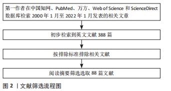
1.1.1 检索人及检索时间 由第一作者在 2022年1月进行检索。
1.1.2 检索文献时限 检索 2000-2022年发表的文献。
1.1.3 检索数据库 PubMed,Web of Science,ScienceDirect,中国知网和万方数据库。
1.1.4 检索词 以“Piezoelectric effect,Piezoelectric materials,Piezoelectric ceramic,Piezoelectric polymers,Osteogenesis,Bone tissue engineering,Scaffolds,Bone defect,energy harvester”为英文检索词;以“压电效应、压电材料、压电陶瓷、压电聚合物、成骨、骨组织工程、细胞支架、骨缺损、能量采集器”为中文检索词。
1.1.5 检索文献类型 检索文献类型 包括研究原著、综述、述评、经验交流、病例报告和荟萃分析。
1.1.6 手工检索情况 无。
1.1.7 检索策略 各检索词之间以 AND 或 OR 逻辑关系连接,具体检索策略见图1。

1.1.8 检索文献量 初检共纳入 388 篇英文文献,20篇中文文献。
1.2 入组标准
1.2.1 纳入标准 ①关于压电材料在骨修复中的研究进展文献;②原创性高、观点明确且论据可靠的高水平文章。
1.2.2 排除标准 ①会议论文及短篇报道;②不符合研究方向的文献;③重复性文献。
1.3 文献质量评估 通过计算机检索,共检索388篇参考文献,按入选标准进行人工筛选,排除不符合研究主题内容及重复的文献。
1.4 数据的提取 首先查看所有文章的标题,摘要,引言和讨论部分,不能判断文献质量的则阅读全文,剔除重复文献,文献内容包括压电材料在骨修复的研究进展。最终共纳入88篇文献,见图2。
3.2 作者综述区别于他人他篇的特点 该综述从压电的角度介绍了骨的压电效应,说明压电材料促进骨修复的可行性以及压电材料应用于骨组织工程的研究进展,细致分析了各种材料的可行性与优缺点,并对压电材料促进成骨潜在的机制做一总结叙述,并为压电材料应用于骨组织工程提供了新的思路。
3.3 综述的局限性 压电材料促进成骨的机制尚不十分清楚,可能的潜在机制与猜想并未全部纳入而仅仅收录了广泛认可的机制,还有一些具有压电效应的生物活性材料如氮化镓、氮化铝镓等限于篇幅限制无法全面介绍。
3.4 综述的重要意义 压电材料与天然骨组织具有相似的压电性能,可以在不借助外接电源与电极的情况下为缺损组织提供良好的电化学微环境。随着研发技术的进步,各式各样的压电材料通过改良与修饰变得愈发契合骨缺损修复的要求,为组织工程技术转化至临床提供新的方向。如何将压电材料的性能与组织修复的机制相结合,如何针对不同病情不同需求的患者提供更加有效的材料将在未来的研究中不断完善。
3.5 课题专家组对未来的建议 随着人口老龄化以及创伤、肿瘤等因素所致难治性骨缺损的患者激增,骨移植手术与骨植入物的需求急需解决。虽然自体骨移植是治疗骨缺损的金标准,然而自体骨获取数量有限难以填补大段骨缺损区域的问题催生了骨组织工程的诞生。骨组织工程以生物支架为载体,将干细胞与药物填补于骨缺损处以促进骨的修复,而研发合适的支架成为了骨组织工程的热点问题。然而,制造具有足够机械强度的、可模拟骨组织微环境且可生物降解的支架并非易事。压电材料的出现为骨组织工程提供了新的思路,压电材料自身可将生理负荷转化为电信号以模拟细胞微环境是自身的优势,研究也表明借助于压电材料可加速骨的修复。然而压电材料自身也存在缺陷,因此将压电材料与其他骨植入材料相结合而形成的复合材料可在保留材料压电性的同时,克服压电材料自身的不足。未来压电复合材料的研发应通过将生物学、工程学、材料学多学科相结合,加强临床医生与材料工程师的密切交流的多学科合作方式,以克服当前生物支架的主要缺陷,提高支架转化为临床应用的效率,以达到改善患者医疗保健和生活质量的目的。
文题释义:
压电材料:某些在受到外力的作用而变形时,其内部会产生极化并使其表面产生电荷现象的晶体材料。骨作为一种天然的压电材料,通过将受到的应力转化为生物电信号,调节骨生长,结构重塑与修复。
细胞外基质:在多细胞有机体中,细胞周围由多种大分子组成的复杂网络。骨组织细胞外基质参与调节细胞黏附、增殖、分化,因此能够良好模拟细胞外基质的生物材料可促进骨修复。
文章从压电的角度详细说明了骨的压电效应以及其在骨修复中的作用原理,说明了压电材料应用于骨组织工程的可行性:为骨细胞创造良好的生物电微环境,具有治疗复杂骨病引起的骨缺损的前景。文章列举了常用的压电材料及其优缺点,压电材料促进骨修复的机制,总结当前压电材料的研究成果与不足,展望了未来压电材料的发展趋势。
| 阅读次数 | ||||||||||||||||||||||||||||||||||||||||||||||||||
|
全文 2316
|
|
|||||||||||||||||||||||||||||||||||||||||||||||||
|
摘要 1352
|
|
|||||||||||||||||||||||||||||||||||||||||||||||||