[1] 张磊,李皓桓,唐金明,等.术中股骨截面后侧皮质线作为股骨前倾角参照在人工全髋关节置换术中的应用[J].临床外科杂志,2018, 26(8):594-597.
[2] 刘建友,贾中伟,牛佳伟,等.构建股骨3D数字化模型提出一种新的股骨颈前倾角测量方法[J].中国组织工程研究,2021,25(24): 3779-3783.
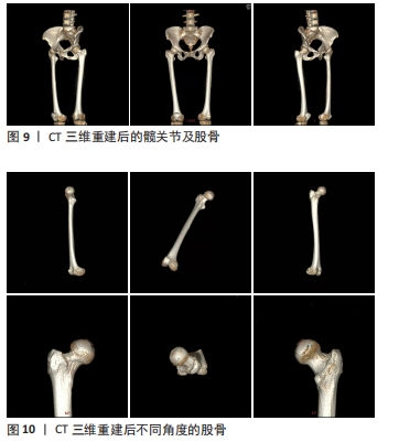
[3] MURPHY SB, SIMON SR, KIJEWSKI PK, et al. Femoral anteversion. J Bone Joint Surg Am. 1987;69(8):1169-1176.
[4] 郑超,马信龙.股骨颈前倾角的测量方法及生物力学[J].中国中西医结合外科杂志,2008,14(3):300-302.
[5] 骆巍,马信龙.股骨头生物力学、解剖形态与血供[J].中国中西医结合外科杂志,2007,13(1):96-98.
[6] 张义修. 骨代谢的生物力学(之一) [J].中国中西医结合外科杂志, 1998,4(4):64-66.
[7] 张义修. 骨代谢的生物力学(之二) [J].中国中西医结合外科杂志, 1998,4(5):318-320.
[8] RUWE PA, GAGE JR, OZONOFF MB, et al. Clinical determination of femoral anteversion. A comparison with established techniques. J Bone Joint Surg Am. 1992;74(6):820-830.
[9] WEDGE JH, MUNKACSI I, LOBACK D. Anteversion of the femur and idiopathic osteoarthrosis of the hip. J Bone Joint Surg Am. 1989;71(7): 1040-1043.
[10] 张怀瑫,郑靖中,杨玉田.国人股骨颈干角及扭转角的测量统计[J].解剖学报,1982,13(3):262-270.
[11] KINGSLEY PC, OLMSTED KL. A study to determine the angle of anteversion of the neck of the femur. J Bone Joint Surg Am. 1948; 30A(3):745-751.
[12] 杨军林,肖学军,彭成宏,等.单张X线片法与CT法测量股骨颈前倾角的比较[J].中华创伤骨科杂志,2004,6(12):1356-1357,1361.
[13] DUNN DM. Anteversion of the neck of the femur; a method of measurement. J Bone Joint Surg Br. 1952;34-B(2):181-186.
[14] OGATA K, GOLDSAND EM. A simple biplanar method of measuring femoral anteversion and neck-shaft angle. J Bone Joint Surg Am. 1979; 61(6A):846-851.
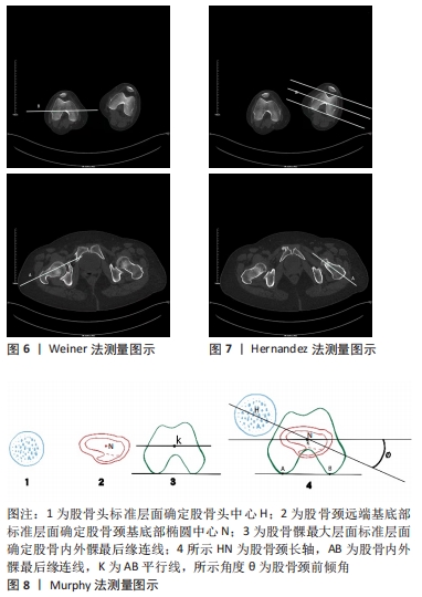
[15] WEINER DS, COOK AJ, HOYT WA JR, et al. Computed tomography in the measurement of femoral anteversion. Orthopedics 1978;1(4):299-306.
[16] HERNANDEZ RJ, TACHDJIAN MO, POZNANSKI AK, et al. CT determination of femoral torsion. AJR Am J Roentgenol. 1981;137(1):97-101.
[17] 杨彦.探讨CT检查的MPR重建图像在角度测量中的运用[J].中国医疗器械信息,2019,25(4):100-102.
[18] 徐高翔,唐佩福.股骨颈前倾角测量方法的回顾与展望[J].解放军医学院学报,2020,41(1):97-100.
[19] 柳达,马瑞雪,吉士俊.股骨颈前倾角的测量及其临床意义[J].中华小儿外科杂志,2002,23(4):78-80.
[20] 马信龙,张清功,马剑雄,等.应用三维重建测量股骨颈前倾角的计算机方法研究[J].生物医学工程与临床,2009,13(5):382-386.
[21] ZHAO JX, SU XY, ZHAO Z, et al. Predicting the optimal entry point for femoral antegrade nailing using a new measurement approach. Int J Comput Assist Radiol Surg. 2015;10(10):1557-1565.
[22] TOMCZAK RJ, GUENTHER KP, RIEBER A, et al. MR imaging measurement of the femoral antetorsional angle as a new technique: comparison with CT in children and adults. AJR Am J Roentgenol. 1997;168(3):791-794.
[23] 张薇,张晶,娄路馨,等.磁共振成像在婴幼儿股骨颈前倾角测量中的应用[J].中医正骨,2013,25(9):29-30,33.
[24] MOULTON A, UPADHYAY SS. A direct method of measuring femoral anteversion using ultrasound. J Bone Joint Surg Br. 1982;64(4):469-472.
[25] TERJESEN T, ANDA S. Femoral anteversion in children measured by ultrasound. Acta Orthop Scand. 1987;58(4):403-407.
[26] CAO J, HU SS, ZHENG HJ, et al. Measurement of femoral neck anteversion of developmental dislocation of hip in children by 3D printing technique. Zhongguo Gu Shang. 2020;33(8):741-744.
[27] BRUNNER A, EICHINGER M, HENGG C, et al. A Simple Method for Measurement of Femoral Anteversion-Validation and Assessment of Reproducibility. J Orthop Trauma. 2016;30(8):e273-278.
[28] 顾硕,赵立来.术中利用C型臂确定股骨颈前倾角的价值分析[J].浙江创伤外科,2020,25(6):1122-1124.
[29] 朱求亮,许斌,沈良华,等.激光投射法测量股骨颈扭转角及前倾角[J].解剖学报,2014,45(5):694-697.
[30] 张德光,韩桂全,张秀玲,等.自制仪器术中股骨颈前倾角测量的临床应用[J].中国实验诊断学,2003,7(4):328-329.
[31] 汪轶平,张恒辉,王燎,等.股骨近端解剖参数的自动化三维测量[J].医用生物力学,2016,31(1):1-7.
[32] CHEN F, LIU J, ZHAO Z, et al. Three-Dimensional Feature-Enhanced Network for Automatic Femur Segmentation. IEEE J Biomed Health Inform. 2019;23(1):243-252.
[33] 柳达,马瑞雪,吉士俊.利用螺旋CT的三维重建技术测量股骨颈前倾角[J].中华小儿外科杂志,2002,23(6):46-50.
[34] 程龙,吉振华.老年股骨颈骨折全髋关节置换术治疗体会[J]. 河南外科学杂志,2016,22(1):96-97.
[35] 迟方舟,张涛,符诗坚,等.国产股骨柄假体在前路髋关节置换中的应用[J].中华关节外科杂志(电子版),2021,15(2):243-247.
[36] 梅晓亮,陈燕,朱伟,等.生物型加长柄在全髋关节置换股骨侧翻修术中的应用[J].中国骨与关节损伤杂志,2021,36(8):794-797.
[37] 马云青,张洪.学习曲线期直接前入路和后外侧入路全髋关节置换术假体位置的比较研究[J].中国骨与关节杂志,2021,10(11):851-856.
[38] 李媛,张恩龙,李文娟,等.人工智能在骨肌系统影像领域的研究进展[J].中国医学科学院学报,2020,42(2):242-246.
[39] 吴东,刘星宇,张逸凌,等.人工智能辅助全髋关节置换术三维规划系统的研发及临床应用研究[J].中国修复重建外科杂志,2020, 34(9):1077-1084.
[40] 张莺.人工智能在医学影像学中的应用[J].中国医疗器械信息,2021, 27(9):64-65.
[41] 郭宇,冯德宏,王凌,等. 3D打印技术在髋关节置换中的应用及价值[J].中国组织工程研究,2020,24(12):1962-1968.
[42] 徐征宇,杜俊炜,姜瑶,等.全髋关节置换术术前模板测量与规划研究进展[J].中华关节外科杂志(电子版),2021,15(1):83-91.
[43] HUPPERTZ A, RADMER S, ASBACH P, et al. Computed tomography for preoperative planning in minimal-invasive total hip arthroplasty: radiation exposure and cost analysis. Eur J Radiol. 2011;78(3):406-413.
|