中国组织工程研究 ›› 2023, Vol. 27 ›› Issue (1): 54-58.doi: 10.12307/2022.998

• 牙髓及牙周膜干细胞 Dental pulp and periodontal ligament stem cells • 上一篇    下一篇

人牙周膜干细胞来源外泌体干预成骨细胞MC3T3-E1的增殖和分化

兰  倩1,辜仰聪2,肖  欣1,毕雪婷1,李  娜3   

  1. 佛山科学技术学院附属口腔医院·佛山市口腔医院,1牙周病科,2口腔颌面外科,广东省佛山市   528099;3佛山科学技术学院医学院,广东省佛山市   528099
  • 收稿日期:2022-01-10 接受日期:2022-02-28 出版日期:2023-01-08 发布日期:2022-06-06
  • 通讯作者: 兰倩,硕士,副主任医师,佛山科学技术学院附属口腔医院·佛山市口腔医院牙周粘膜科,广东省佛山市 528099
  • 作者简介:兰倩,女,1980年生,四川省成都市人,汉族,2005年四川大学华西口腔医学院毕业,硕士,副主任医师,主要从事牙周病学研究。
  • 基金资助:
    广东省基础与应用研究基金粤佛联合基金(2019A1515110013),项目负责人:李娜

Human periodontal ligament stem cells-derived exosomes interfere with the proliferation and differentiation of MC3T3-E1 cells

Lan Qian1, Gu Yangcong2, Xiao Xin1, Bi Xueting1, Li Na3   

  1. 1Department of Periodontics, 2Department of Oral and Maxillofacial Surgery, Foshan Stomatology Hospital, School of Stomatology and Medicine, Foshan University, Foshan 528099, Guangdong Province, China; 3Medical College, Foshan University Foshan 528099, Guangdong Province, China
  • Received:2022-01-10 Accepted:2022-02-28 Online:2023-01-08 Published:2022-06-06
  • Contact: Lan Qian, Master, Associate chief physician, Department of Periodontics, Foshan Stomatology Hospital, School of Stomatology and Medicine, Foshan University, Foshan 528099, Guangdong Province, China
  • About author:Lan Qian, Master, Associate chief physician, Department of Periodontics, Foshan Stomatology Hospital, School of Stomatology and Medicine, Foshan University, Foshan 528099, Guangdong Province, China
  • Supported by:
    Guangdong Fundamental and Applied Research Fund Yue-Fo Joint Fund, No. 2019A1515110013 (to LN)

摘要:

文题释义:
牙周膜干细胞来源外泌体:在牙周膜干细胞体外培养过程中,分泌到培养基中的一种直径在30-120 nm间的纳米级囊泡物,内含有蛋白质、脂质、DNA、编码和非编码RNA及代谢物等生物学活性物质。
MC3T3-E1细胞:小鼠胚胎成骨细胞前体细胞系,形态为成纤维细胞样,有多个亚克隆,可以作为体外研究成骨细胞分化的良好模型。

背景:人牙周膜干细胞具有较强的成骨分化能力,人牙周膜干细胞来源外泌体作为牙周膜干细胞分泌的主要成分,对成骨细胞MC3T3-E1增殖和成骨分化的影响尚不明确。
目的:探讨人牙周膜干细胞来源外泌体对MC3T3-E1细胞增殖和分化的影响。
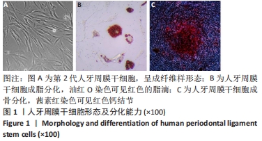
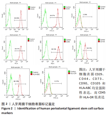
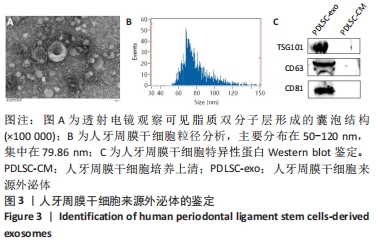
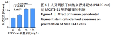
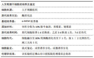
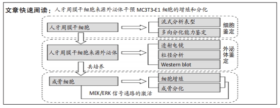
方法:采用酶消化法分离及培养人牙周膜干细胞,超速离心法提取人牙周膜干细胞来源外泌体,通过透射电镜、粒径分析及Western blot方法对人牙周膜干细胞来源外泌体进行鉴定;CCK8法检测不同质量浓度人牙周膜干细胞来源外泌体对MC3T3-E1细胞增殖的影响,茜素红染色观察100 mg/L人牙周膜干细胞来源外泌体对MC3T3-E1细胞成骨矿化的影响,Western blot检测100 mg/L人牙周膜干细胞来源外泌体干预前后MC3T3-E1细胞内MEK和ERK的磷酸化水平。
结果与结论:①透射电镜观察可见外泌体为脂质双分子层形成的囊泡结构,粒径检测显示外泌体直径分布在50-120 nm,集中在
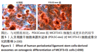
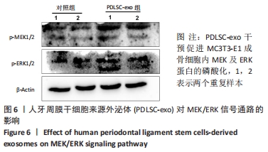
79.86 nm,Western blot检测结果显示提取的外泌体中含有CD81,CD63,TSG101的表达;②与对照组相比,人牙周膜干细胞来源外泌体对MC3T3-E1细胞的增殖具有促进作用,且作用呈剂量依赖性;③与对照组相比,人牙周膜干细胞来源外泌体组MC3T3-E1细胞能够形成更多的钙结节;与对照组相比,人牙周膜干细胞来源外泌体组MC3T3-E1细胞内p-MEK及p-ERK蛋白表达量升高;④结果表明,人牙周膜干细胞来源外泌体可以显著促进MC3T3-E1增殖和成骨分化,推测可能与其激活MEK/ERK信号通路有关。
缩略语:人牙周膜干细胞来源外泌体:periodontal ligament stem cells derived exosomes,PDLSC-exo

https://orcid.org/0000-0002-2010-6836 (兰倩)

中国组织工程研究杂志出版内容重点:干细胞;骨髓干细胞;造血干细胞;脂肪干细胞;肿瘤干细胞;胚胎干细胞;脐带脐血干细胞;干细胞诱导;干细胞分化;组织工程

关键词: 牙周膜干细胞, 外泌体, 成骨细胞, MC3T3-E1细胞, 增殖, 分化

Abstract: BACKGROUND: Human periodontal ligament stem cells had a strong osteogenic ability, but the effect of human periodontal ligament stem cells-derived exosomes on proliferation and osteogenic differentiation of MC3T3-E1 is not clear. 
OBJECTIVE: To explore the effect of human periodontal ligament stem cells-derived exosomes on the proliferation and differentiation of MC3T3-E1 cells.
METHODS: Human periodontal ligament stem cells were isolated and cultured by enzyme digestion. Human periodontal ligament stem cells-derived exosomes were extracted by ultracentrifugation, and identified by transmission electron microscopy, nanoparticle tracking analysis and western blot assay. Effect of different mass concentrations of human periodontal ligament stem cells-derived exosomes on proliferation of MC3T3-E1 cells was detected by CCK-8 assay. The effect of 100 mg/L human periodontal ligament stem cells-derived exosomes on osteogenic mineralization of MC3T3-E1 cells was observed by alizarin red staining. Western blot assay was used to detect the phosphorylation levels of MEK and ERK in MC3T3-E1 cells before and after the intervention of 100 mg/L human periodontal ligament stem cells-derived exosomes.  
RESULTS AND CONCLUSION: (1) Transmission electron microscopy showed that exosomes were vesicles formed by lipid bilayer. Nanoparticle tracking analysis showed that exosomes were 50-120 nm in diameter and concentrated at 79.86 nm. Western blot assay showed that the exosomes contained expression levels of CD81, CD63 and TSG101. (2) Compared with the control group, human periodontal ligament stem cells-derived exosomes could promote the proliferation of MC3T3-E1 cells in a dose-dependent manner. (3) Compared with the control group, MC3T3-E1 cells were able to form many calcium nodules in the human periodontal ligament stem cell-derived exosome group. Compared with the control group, protein expression of p-MEK and p-ERK increased in MC3T3-E1 cells of the human periodontal ligament stem cell-derived exosome group. (4) It is indicated that human periodontal ligament stem cells-derived exosomes can promote the proliferation and osteogenic differentiation of MC3T3-E1 cells, which may be associated with the activation of MEK/ERK signaling pathway. 

Key words: periodontal ligament stem cell, exosome, osteoblast, MC3T3-E1 cell, proliferation, differentiation

中图分类号: