[1] GOEDEKE L, PERRY RJ, SHULMAN GI. Emerging Pharmacological Targets for the Treatment of Nonalcoholic Fatty Liver Disease, Insulin Resistance, and Type 2 Diabetes. Ann Rev Pharmacol. 2019;59(1):65-87.
[2] KLISIC A, ISAKOVIC A, KOCIC G, et al. Relationship between Oxidative Stress, Inflammation and Dyslipidemia with Fatty Liver Index in Patients with Type 2 Diabetes Mellitus. Exp Clin Endocrinol Diabetes. 2018;126(6):371-378.
[3] FRANCESCONI C, NIEBAUER J, HABER P, et al. Lebensstil: körperliche Aktivität und Training in der Prävention und Therapie des Typ 2 Diabetes mellitus (Update 2019) [Lifestyle: physical activity and training as prevetion and therapy of type 2 diabetes mellitus (Update 2019)]. Wien Klin Wochenschr. 2019;131(Suppl 1):61-66.
[4] ELIAS D, PRIGOZIN H, POLAK N, et al. Autoimmune diabetes induced by the beta-cell toxin STZ. Immunity to the 60-kDa heat shock protein and to insulin. Diabetes. 1994;43(8):992-998.
[5] BEDFORD TG, TIPTON CM, WILSON NC, et al. Maximum oxygen consumption of rats and its changes with various experimental procedures. J Appl Physiol Respir Environ Exerc Physiol. 1979;47(6): 1278-1283.
[6] HORNBERGER TA JR, FARRAR RP. Physiological hypertrophy of the FHL muscle following 8 weeks of progressive resistance exercise in the rat. Can J Appl Physiol. 2004;29(1):16-31.
[7] AL-AWAR A, KUPAI K, VESZELKA M, et al. Experimental Diabetes Mellitus in Different Animal Models. J Diabetes Res. 2016;2016:9051426.
[8] GHEIBI S, KASHFI K, GHASEMI A. A practical guide for induction of type-2 diabetes in rat: Incorporating a high-fat diet and streptozotocin. Biomed Pharmacother. 2017;95:605-613.
[9] MEHTA BK, SINGH KK, BANERJEE S. Effect of exercise on type 2 diabetes-associated cognitive impairment in rats. Int J Neurosci. 2019; 129(3):252-263.
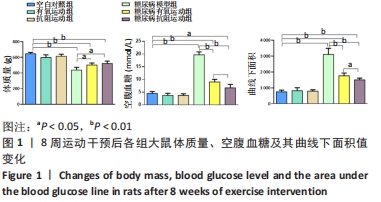
[10] 尚画雨,夏志,张丹,等.8周游泳运动对2型糖尿病大鼠周围神经病变的影响[J].中国病理生理杂志,2014,30(4):719-724.
[11] CHO NH, SHAW JE, KARURANGA S, et al. IDF Diabetes Atlas: Global estimates of diabetes prevalence for 2017 and projections for 2045. Diabetes Res Clin Pract. 2018;138:271-281.
[12] GHASEMI A, JEDDI S. Anti-obesity and anti-diabetic effects of nitrate and nitrite. Nitric Oxide. 2017;70:9-24.
[13] CZECH MP. Insulin action and resistance in obesity and type 2 diabetes. Nat Med. 2017;23(7):804-814.
[14] BARAZZONI R, GORTAN CAPPELLARI G, et al. Insulin resistance in obesity: an overview of fundamental alterations. Eat Weight Disord. 2018;23(2):149-157.
[15] KIRWAN JP, SACKS J, NIEUWOUDT S. The essential role of exercise in the management of type 2 diabetes. Cleve Clin J Med. 2017;84(7 Suppl 1): S15-S21.
[16] YANG D, YANG Y, LI Y, et al. Physical Exercise as Therapy for Type 2 Diabetes Mellitus: From Mechanism to Orientation. Ann Nutr Metab. 2019;74(4):313-321.
[17] AMANAT S, GHAHRI S, DIANATINASAB A, et al. Exercise and Type 2 Diabetes. Adv Exp Med Biol. 2020;1228:91-105.
[18] DONGHUI T, SHUANG B, XULONG L, et al. Improvement of microvascular endothelial dysfunction induced by exercise and diet is associated with microRNA-126 in obese adolescents. Microvasc Res. 2019;123:86-91.
[19] RÖHLING M, HERDER C, RODEN M, et al. Effects of Long-Term Exercise Interventions on Glycaemic Control in Type 1 and Type 2 Diabetes: a Systematic Review. Exp Clin Endocrinol Diabetes. 2016;124(8):487-494.
[20] CHIANG SL, HEITKEMPER MM, HUNG YJ, et al. Effects of a 12-week moderate-intensity exercise training on blood glucose response in patients with type 2 diabetes: A prospective longitudinal study. Medicine (Baltimore). 2019;98(36):e16860.
[21] LI S, LIANG M, PAN Y, et al. Exercise modulates heat shock protein 27 activity in diabetic cardiomyopathy. Life Sci. 2020;243:117251.
[22] MANNUCCI E, BONIFAZI A, MONAMI M. Comparison between different types of exercise training in patients with type 2 diabetes mellitus: A systematic review and network metanalysis of randomized controlled trials. Nutr Metab Cardiovasc Dis. 2021;31(7):1985-1992.
[23] REDDY R, WITTENBERG A, CASTLE JR, et al. Effect of Aerobic and Resistance Exercise on Glycemic Control in Adults With Type 1 Diabetes. Can J Diabetes. 2019;43(6):406-414.e1.
[24] LI S, LIANG M, GAO D, et al. Changes in Titin and Collagen Modulate Effects of Aerobic and Resistance Exercise on Diabetic Cardiac Function. J Cardiovasc Transl Res. 2019;12(5):404-414.
[25] NERY C, MORAES SRA, NOVAES KA, et al. Effectiveness of resistance exercise compared to aerobic exercise without insulin therapy in patients with type 2 diabetes mellitus: a meta-analysis.Braz J Phys Ther. 2017;21(6):400-415.
[26] KARSTOFT K, PEDERSEN BK. Exercise and type 2 diabetes: focus on metabolism and inflammation. Immunol Cell Biol. 2016;94(2):146-150.
[27] JAFARIPOUR S, SEDIGHI S, JOKAR MH, et al. Inflammation, diet, and type 2 diabetes: a mini-review. J Immunoassay Immunochem. 2020; 41(4):768-777.
[28] PEDERSEN BK. Anti-inflammatory effects of exercise: role in diabetes and cardiovascular disease. Eur J Clin Invest. 2017;47(8):600-611.
[29] MEIJNIKMAN AS, GERDES VE, NIEUWDORP M, et al. Evaluating Causality of Gut Microbiota in Obesity and Diabetes in Humans. Endocr Rev. 2018;39(2):133-153.
[30] ZHOU W, CHEN C, CHEN Z, et al. NLRP3: A Novel Mediator in Cardiovascular Disease. J Immunol Res. 2018;2018:5702103.
[31] AN N, GAO Y, SI Z, et al. Regulatory Mechanisms of the NLRP3 Inflammasome, a Novel Immune-Inflammatory Marker in Cardiovascular Diseases. Front Immunol. 2019;10:1592.
[32] HONG P, GU RN, LI FX, et al. NLRP3 inflammasome as a potential treatment in ischemic stroke concomitant with diabetes. J Neuroinflammation. 2019;16(1):121.
[33] KAYAGAKI N, STOWE IB, LEE BL, et al. Caspase-11 cleaves gasdermin D for non-canonical inflammasome signalling. Nature. 2015;526(7575): 666-671.
[34] LI W, CHANG Y, LIANG S, et al. NLRP3 inflammasome activation contributes to Listeria monocytogenes-induced animal pregnancy failure. BMC Vet Res. 2016;12:36.
[35] SHARMA D, KANNEGANTI TD. The cell biology of inflammasomes: Mechanisms of inflammasome activation and regulation. Cell Biol. 2016;213(6):617-629.
[36] MALIK A, KANNEGANTI TD. Inflammasome activation and assembly at a glance. J Cell Sci. 2017;130(23):3955-3963.
[37] DING S, XU S, MA Y, et al. Modulatory Mechanisms of the NLRP3 Inflammasomes in Diabetes. Biomolecules. 2019;9(12):850.
[38] ZHANG W, XU X, KAO R, et al. Cardiac fibroblasts contribute to myocardial dysfunction in mice with sepsis: the role of NLRP3 inflammasome activation. PLoS One. 2014;9(9):e107639.
[39] XIE Y, HUANG Y, LING X, et al. Chemerin/CMKLR1 Axis Promotes Inflammation and Pyroptosis by Activating NLRP3 Inflammasome in Diabetic Cardiomyopathy Rat. Front Physiol. 2020;11:381.
[40] LUO B, HUANG F, LIU Y, et al. NLRP3 Inflammasome as a Molecular Marker in Diabetic Cardiomyopathy. Front Physiol. 2017;8:519.
[41] LI X, LI Z, LI B, et al. Klotho improves diabetic cardiomyopathy by suppressing the NLRP3 inflammasome pathway. Life Sci. 2019;234: 116773.
[42] VANDANMAGSAR B, YOUM YH, RAVUSSIN A, et al. The NLRP3 inflammasome instigates obesity-induced inflammation and insulin resistance. Nat Med. 2011;17(2):179-188.
[43] WANG Q, HU J, LIU Y, et al. Aerobic Exercise Improves Synaptic-Related Proteins of Diabetic Rats by Inhibiting FOXO1/NF-κB/NLRP3 Inflammatory Signaling Pathway and Ameliorating PI3K/Akt Insulin Signaling Pathway. J Mol Neurosci. 2019;69(1):28-38.
[44] LI J, LIU Y, LIU B, et al. Mechanisms of Aerobic Exercise Upregulating the Expression of Hippocampal Synaptic Plasticity-Associated Proteins in Diabetic Rats. Neural Plast. 2019;2019:7920540.
[45] MARDARE C, KRÜGER K, LIEBISCH G, et al. Endurance and Resistance Training Affect High Fat Diet-Induced Increase of Ceramides, Inflammasome Expression, and Systemic Inflammation in Mice. J Diabetes Res. 2016;2016:4536470.
[46] YANG W, LIU L, WEI Y, et al. Exercise suppresses NLRP3 inflammasome activation in mice with diet-induced NASH: a plausible role of adropin. Lab Invest. 2021;101(3):369-380.
[47] HONG J, PARK E, LEE J, et al. Exercise training mitigates ER stress and UCP2 deficiency-associated coronary vascular dysfunction in atherosclerosis. Sci Rep. 2021;11(1):15449.
[48] ZHUGE DL, JAVAID HMA, SAHAR NE, et al. Fibroblast growth factor 2 exacerbates inflammation in adipocytes through NLRP3 inflammasome activation. Arch Pharm Res. 2020;43(12):1311-1324.
[49] KHAKROO ABKENAR I, RAHMANI-NIA F, LOMBARDI G. The effects of acute and chronic aerobic activity on the signaling pathway of the inflammasome NLRP3 complex in young men. Medicina. 2019;55(4): 105.
|