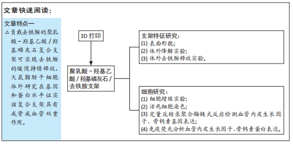
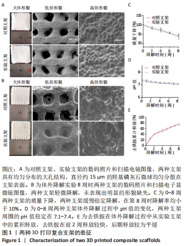
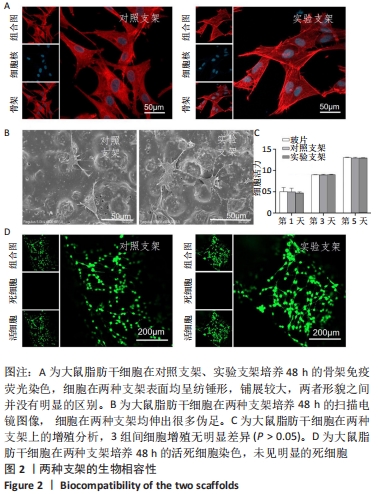
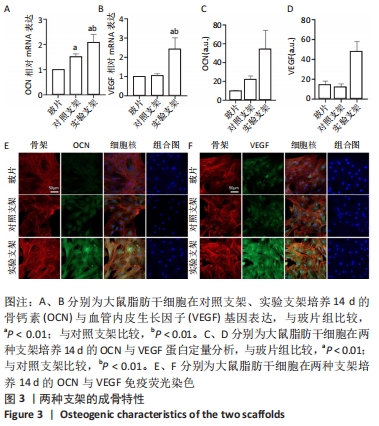
[1] LIU WC, CHEN S, ZHENG L, et al. Angiogenesis Assays for the Evaluation of Angiogenic Properties of Orthopaedic Biomaterials - A General Review. Adv Healthc Mater. 2017;6(5):1600434.
[2] SARAN U, GEMIBU PIPERNI S, CHATTERJEE S, et al. Role of angiogenesis in bone repair. Arch Biochem Biophys. 2014;1(561):109-117.
[3] HAUSMAN MR, SCHAFFLER MB, MAJESKA RJ, et al. Prevention of fracture healing in rats by an inhibitor of angiogenesis. Bone. 2001; 29(6):560-564.
[4] HUANG JH, LIN D, WEI ZY, et al. Parathyroid Homone Derivative with Reduced Osteoclastic Activity Promoted Bone Regeneration via Synergistic Bone Remodeling and Angiogenesis. Small. 2020;16(6): 1905876.
[5] DANHIER F, ANSORENA E, SILVA JM, et al. PLGA-based nanoparticles: an overview of biomedical applications. J Control Release. 2012; 161(2): 505-522.
[6] WENZ A, BORCHERS KIRSTEN, TOVAR GEM, et al. Bone matrix production in hydroxyapatite-modified hydrogels suitable for bone bioprinting. Biofabrication. 2017;9(4):044103.
[7] BENDTSEN ST, QUINNELL SP, WEI M, et al. Development of a novel alginate-polyvinyl alcohol-hydroxyapatite hydrogel for 3D bioprinting bone tissue engineered scaffolds. J Biomed Mater Res A. 2017;105(5): 1457-1468.
[8] YOU F, CHEN XB, COOPER DML, et al. Homogeneous hydroxyapatite/alginate composite hydrogel promotes calcified cartilage matrix deposition with potential for three-dimensional bioprinting. Biofabrication. 2018;11(1):015015.
[9] JAKUS AE, SHAH RN. Multi and mixed 3D-printing of graphene-hydroxyapatite hybrid materials for complex tissue engineering. J Biomed Mater Res A. 2017;105(1):274-283.
[10] SZCZES A, HOLYSZ L, CHIBOWSKI E. Synthesis of hydroxyapatite for biomedical applications. Adv Colloid Interfac. 2017;249:321-330.
[11] SATTARY M, RAFIENIA M, KAZEMI M, et al. Promoting effect of nano hydroxyapatite and vitamin D3 on the osteogenic differentiation of human adipose-derived stem cells in polycaprolactone/gelatin scaffold for bone tissue engineering. Mat Sci Eng C-Mater. 2019;97:141-155.
[12] GAO L, HUANG ZY, YAN SF, et al. Sr-HA-graft-Poly(gamma-benzyl-l-glutamate) Nanocomposite Microcarriers: Controllable Sr(2+) Release for Accelerating Osteogenenisis and Bony Nonunion Repair. Biomacromolecules. 2017;18(11):3742-3752.
[13] MARX RE, SAWATARI Y, FORTIN M, et al. Bisphosphonate-induced exposed bone (osteonecrosis/osteopetrosis) of the jaws: risk factors, recognition, prevention, and treatment. J Oral Maxillofac Surg. 2005; 63(11):1567-1575.
[14] MONDAL S, DOROZHKIN SV, PAL U, et al. Recent progress on fabrication and drug delivery applications of nanostructured hydroxyapatite. Wires Nanomed Nanobi. 2018;10(4):e1504.
[15] GENTILE P, CHIONO V, CARMAGNOLA I, et al. An overview of poly(lactic-co-glycolic) acid (PLGA)-based biomaterials for bone tissue engineering. Int J Mol Sci. 2014;15(3):3640-3659.
[16] XIE XH, WANG XL, ZHANG Z, et al. Structural and degradation characteristics of an innovative porous PLGA/TCP scaffold incorporated with bioactive molecular icaritin. Biomed Mater. 2010;5(5):054109.
[17] QIN L, YAO D, ZHENG LZ, et al. Phytomolecule icaritin incorporated PLGA/TCP scaffold for steroid-associated osteonecrosis: Proof-of-concept for prevention of hip joint collapse in bipedal emus and mechanistic study in quadrupedal rabbits. Biomaterials. 2015;59:125-143.
[18] WANG XL, XIE XH, ZHANG G, et al. Exogenous phytoestrogenic molecule icaritin incorporated into a porous scaffold for enhancing bone defect repair. J Orthop Res. 2013;31(1):164-172.
[19] HOLDEN P, NAIR LS. Deferoxamine: An Angiogenic and Antioxidant Molecule for Tissue Regeneration. Tissue Eng Part B-Re. 2019;25(6): 461-470.
[20] TCHANQUE-FOSSUO CN, DAHLE SE, BUCHMAN SR, et al. Deferoxamine: potential novel topical therapeutic for chronic wounds. Brit J Dermatol. 2017;176(4):1056-1059.
[21] DUSCHER D, TROTSYUK AA, MAAN ZN, et al. Optimization of transdermal deferoxamine leads to enhanced efficacy in healing skin wounds. J Control Release. 2019;308:232-239.
[22] QAYOOM A, ANEESHA VA, ANAGHA S, et al. Lecithin-based deferoxamine nanoparticles accelerated cutaneous wound healing in diabetic rats. Eur J Pharmacol. 2019;858:172478.
[23] YAN YF, CHEN H, ZHANG HB, et al. Vascularized 3D printed scaffolds for promoting bone regeneration. Biomaterials. 2019;190-191:97-110.
[24] LI HH, LUO BH, WEN W, et al. Deferoxamine immobilized poly(D,L-lactide) membrane via polydopamine adhesive coating: The influence on mouse embryo osteoblast precursor cells and human umbilical vein endothelial cells. Mat Sci Eng C-Mater. 2017;70:701-709.
[25] RAN QC, YU YL, CHEN WZ, et al. Deferoxamine loaded titania nanotubes substrates regulate osteogenic and angiogenic differentiation of MSCs via activation of HIF-1alpha signaling. Mat Sci Eng C-Mater. 2018;91: 44-54.
[26] JIA P, CHEN H, KANG H, et al. Deferoxamine released from poly(lactic-co-glycolic acid) promotes healing of osteoporotic bone defect via enhanced angiogenesis and osteogenesis. J Biomed Mater Res A. 2016; 104(10):2515-2527.
[27] WEN Y, XUN S, HAOYE M, et al. 3D printed porous ceramic scaffolds for bone tissue engineering: a review. Biomater Sci-UK. 2017;5(9):1690-1698.
[28] VENKATESAN J, BHATNAGAR I, MANIVASAGAN P, et al. Alginate composites for bone tissue engineering: A review. Int J Biol Macromol. 2015;72:269-281.
[29] LECOMTE A, GAUTIER H, BOULER JM, et al. Biphasic calcium phosphate: a comparative study of interconnected porosity in two ceramics. J Biomed Mater Res B. 2008;84(1):1-6.
[30] PEI X, MA L, ZHANG BQ, et al. Creating hierarchical porosity hydroxyapatite scaffolds with osteoinduction by three-dimensional printing and microwave sintering. Biofabrication. 2017;9(4):045008.
[31] WANG XJ, LOU T, ZHAO WH, et al. The effect of fiber size and pore size on cell proliferation and infiltration in PLLA scaffolds on bone tissue engineering. J Biomater Appl. 2016;30(10):1545-1551.
[32] LAI YX, CAO HJ, WANG XL, et al. Porous composite scaffold incorporating osteogenic phytomolecule icariin for promoting skeletal regeneration in challenging osteonecrotic bone in rabbits. Biomaterials. 2018;153:1-13.
[33] CHEN SH, LEI M, XIE XH, et al. PLGA/TCP composite scaffold incorporating bioactive phytomolecule icaritin for enhancement of bone defect repair in rabbits. Acta Biomater. 2013;9(5):6711-6722.
[34] XIE XH, WANG XL, ZHENG G, et al. Biofabrication of a PLGA-TCP-based porous bioactive bone substitute with sustained release of icaritin. J Tissue Eng Regen M. 2015;9(8):961-972.
|