中国组织工程研究 ›› 2022, Vol. 26 ›› Issue (28): 4441-4445.doi: 10.12307/2022.295

• 组织工程骨材料 tissue-engineered bone • 上一篇    下一篇

柚皮苷-壳聚糖/羟基磷灰石复合支架修复大鼠颅骨缺损

卢育南1,张信照1,林斌斌1,徐  敢2,陈景帝2,陈顺有1   

  1. 1厦门大学附属福州第二医院小儿骨科,福建省福州市  350007;2福州大学生物和医药技术研究院,福建省福州市  350002
  • 收稿日期:2020-12-04 接受日期:2021-01-16 出版日期:2022-10-08 发布日期:2022-03-17
  • 通讯作者: 陈顺有,副主任医师,医学博士,厦门大学附属福州第二医院小儿骨科,福建省福州市 350007
  • 作者简介:卢育南,男,1990年生,福建省漳州市人,汉族,主治医师,医学硕士,主要从事儿童骨科研究。
  • 基金资助:
    福州市科技计划项目(2018-S-101-3),项目负责人:卢育南

Naringin-chitosan/hydroxyapatite composite scaffold in repair of rat skull defect

Lu Yunan1, Zhang Xinzhao1, Lin Binbin1, Xu Gan2, Chen Jingdi2, Chen Shunyou1   

  1. 1Department of Pediatric Orthopedics, Fuzhou Second Hospital Affiliated to Xiamen University, Fuzhou 350007, Fujian Province, China; 2Institute of Biological and Medical Technology, Fuzhou University, Fuzhou 350002, Fujian Province, China
  • Received:2020-12-04 Accepted:2021-01-16 Online:2022-10-08 Published:2022-03-17
  • Contact: Chen Shunyou, MD, Associate chief physician, Department of Pediatric Orthopedics, Fuzhou Second Hospital Affiliated to Xiamen University, Fuzhou 350007, Fujian Province, China
  • About author:Lu Yunan, Master, Attending physician, Department of Pediatric Orthopedics, Fuzhou Second Hospital Affiliated to Xiamen University, Fuzhou 350007, Fujian Province, China
  • Supported by:
    the Fuzhou Science and Technology Plan Project, No. 2018-S-101-3 (to LYN)

摘要:

文题释义:
柚皮苷:是由水龙骨科植物槲蕨根茎(骨碎补)中提取的主要有效成分,能够促进成骨细胞增殖、促进血管生成、抑制破骨细胞生长的功能。
壳聚糖/羟基磷灰石复合支架:将一定钙磷比的羟基磷灰石前体液与壳聚糖溶液在碱性条件下原位结晶、冷冻干燥后可以获得多孔的支架结构,可作为人工骨用于骨缺损的修复治疗。

背景:骨碎补的有效成分柚皮苷具有补肝肾强筋骨的传统功效,能增加骨痂厚度,提高骨折愈合质量。
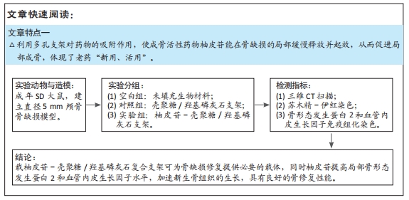
目的:探究载中药骨碎补有效成分柚皮苷壳聚糖/羟基磷灰石复合支架的骨传导和骨诱导性能。
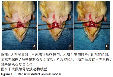
方法:将一定钙磷比的羟基磷灰石前体液与含柚皮苷的壳聚糖溶液在碱性条件下原位结晶、冷冻干燥,获得柚皮苷-壳聚糖/羟基磷灰石多孔支架。将15只成年SD大鼠随机分成空白组(n=5)、对照组(n=5)和实验组(n=5),建立直径5 mm颅骨骨缺损模型,空白组未填充生物材料,对照组填充壳聚糖/羟基磷灰石支架,实验组填充柚皮苷-壳聚糖/羟基磷灰石复合支架。术后4周取材,CT扫描观察颅骨修复情况,苏木精-伊红染色观察颅骨修复的形态学,骨形态发生蛋白2和血管内皮生长因子免疫组化染色后观察缺损区域局部成骨活性因子的表达。
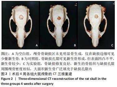
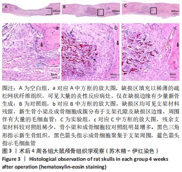
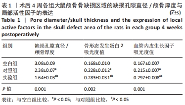
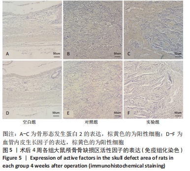
结果与结论:①CT扫描显示,空白组大鼠颅骨未见明显骨生成,仅在缺损边缘可见少量新生骨;对照组于缺损孔隙可见新生骨形成,新生骨较少;实验组骨缺损修复良好,新生骨组织与缺损孔隙周围颅骨密度相似,大面积新生骨广泛填充了缺损孔隙。②苏木精-伊红染色显示,空白组缺损区填充以稀薄的疏松网状纤维组织,可见大量炎性反应病灶,仅在缺损边缘有少量新骨生成;对照组与实验组缺损区均可见支架材料残留,新生骨小梁及成骨细胞成簇分布于支架孔隙及缺损区边缘,周围伴有大量的毛细血管,其中以实验组表现出更强的新骨爬行替代能力。③免疫组化染色显示,实验组局部成骨活性因子骨形态发生蛋白2 和血管内皮生长因子的表达高于对照组、空白组(P < 0.05)。④结果表明,柚皮苷-壳聚糖/羟基磷灰石复合支架可为骨缺损修复提供必要的载体,柚皮苷可创造局部成骨微环境、加速新生骨组织的生长,具有良好的骨修复性能。

https://orcid.org/0000-0002-6594-9917 (卢育南) 

中国组织工程研究杂志出版内容重点:生物材料;骨生物材料口腔生物材料纳米材料缓释材料材料相容性组织工程

关键词: 柚皮苷, 壳聚糖, 羟基磷灰石, 复合支架, 骨缺损, 成骨性能

Abstract: BACKGROUND: Naringin, the active ingredient of Rhizoma Drynariae, has the traditional effect of strengthening the liver and kidney and strengthening the bones and muscles, increasing the thickness of the callus and improving the quality of fracture healing.
OBJECTIVE: To explore the bone conduction and bone induction properties of naringin-chitosan/hydroxyapatite composite scaffolds.  
METHODS: The hydroxyapatite precursor solution with a certain calcium to phosphorus ratio and the chitosan solution containing naringin were crystallized in situ under alkaline conditions and freeze-dried to obtain a naringin-chitosan/hydroxyapatite porous scaffold. A total of 15 SD rats were randomly divided into blank group (n=5), control group (n=5) and experimental group (n=5). The 5 mm-diameter skull bone defect models were established by drilling holes. Biomaterials were not filled in the blank group. Chitosan/hydroxyapatite scaffolds were filled in the control group; and naringin-chitosan/hydroxyapatite scaffolds were filled in the experimental group. At 4 weeks after surgery, CT scans were performed to observe the skull repair. Hematoxylin-eosin staining was used to observe morphological differences. Immunohistochemical staining of bone morphogenetic protein 2 and vascular endothelial growth factor was performed to observe the expression of local osteogenic active factors in the defect areas. 
RESULTS AND CONCLUSION: (1) CT scan showed that no obvious osteogenesis was seen in the skull of rats in the blank group; only a small amount of new bone was seen at the edge of the defect. In the control group, new bone formation could be seen in the defect pores, and there was less new bone. In the experimental group, the bone defect was well repaired; the density of new bone tissue and the skull around the defect pores were similar, and a large area of new bone widely filled the defect pores. (2) Hematoxylin-eosin staining showed that the defect area in the blank group was filled with thin loose reticular fibrous tissue, and a large number of inflammatory response lesions were seen, and only a small amount of new bone was formed at the edge of the defect. In the control group and the experimental group, residual scaffold material, new bone trabecula and osteoblasts were seen in the defect area; osteoblasts were distributed in clusters in the pores of the scaffold and the edges of the defect area, surrounded by a large number of capillaries. Among them, the experimental group showed a stronger ability of new bone growth. (3) Immunohistochemical staining showed that the expression levels of local osteogenic activity factors bone morphogenetic protein 2 and vascular endothelial growth factor in the experimental group were higher than those in the control and blank groups (P < 0.05). (4) The results conclude that naringin-chitosan/hydroxyapatite composite scaffolds can provide necessary carrier for bone defect repair. Naringin can create local osteogenic microenvironment, accelerate the growth and mineralization of new bone tissue, and have good bone repair performance.

Key words: naringin, chitosan, hydroxyapatite, composite scaffold, bone defect, osteogenesis performance

中图分类号: