[1]VERGROESEN PP, KINGMA I, EMANUEL KS, et al. Mechanics and biology in intervertebral disc degeneration: a vicious circle. Osteoarthritis Cartilage. 2015;23(7): 1057-1070.
[2]KERMANI HR, HOBOUBATI H, ESMAEILI-MAHANI S, et al. Induction of intervertebral disc cell apoptosis and degeneration by chronic unpredictable stress. J Neurosurg Spine. 2014;20(5): 578-584.
[3]LI N, GAO Q, ZHOU W, et al. MicroRNA-129-5p affects immune privilege and apoptosis of nucleus pulposus cells via regulating FADD in intervertebral disc degeneration. Cell Cycle. 2020;11(1): 1-16.
[4]SHEN J, FANG J, HAO J, et al. SIRT1 Inhibits the Catabolic Effect of IL-1β Through TLR2/SIRT1/NF-κB Pathway in Human Degenerative Nucleus Pulposus Cells. Pain Phys. 2016;19(1): E215-E216.
[5]TONG P, PENG QH, GU LM, et al. LncRNA-MEG3 alleviates high glucose induced inflammation and apoptosis of retina epithelial cells via regulating miR-34a/SIRT1 axis. Exp Mol Pathol. 2019;107(1): 102-109.
[6] 孙中仪, 晏鹏, 王圣杰,等. 长链非编码RNA在人退变椎间盘组织中的基因表达谱[J]. 中华医学杂志, 2017, 97(33):2582-2586.
[7] 王毅峰. LncRNA在椎间盘退变过程中调控髓核细胞凋亡的功能及其机制研究[D].上海:第二军医大学, 2016.
[8]Bai X, Guo X, Zhang F, et al. Resveratrol Combined with 17β-Estradiol Prevents IL-1β Induced Apoptosis in Human Nucleus Pulposus Via The PI3K/AKT/Mtor and PI3K/AKT/GSK-3β Pathway. J Invest Surg. 2020; 10(1): 1-8.
[9] 陈江, 刘志超, 张帆,等. 身痛逐瘀汤对人髓核细胞模型PI3K/Akt信号通路Bad、Caspase-9、GSK-3β表达的影响[J]. 中国中医药信息杂志, 2019, 26(9):48-53.
[10] 鲁花, 于露, 甄欢欢,等. 骨形态发生蛋白9激活PI3K/Akt信号通路抑制退变髓核细胞的炎症反应和凋亡[J]. 第三军医大学学报, 2016, 38(18):2047-2052.
[11]吴瑞凯, 齐义营, 茹选良, 等. 血清饥饿对大鼠椎间盘髓核细胞凋亡的影响及其机制研究[J]. 中华全科医学, 2016, 14(6): 889-892.
[12]张海丹, 李培武, 张玲. 五味子乙素通过调控p38MAPK信号通路对IL-1β诱导的髓核细胞凋亡的影响[J]. 医学分子生物学杂志, 2019, 16(2): 119-124.
[13]郑文凯, 黄智, 达逸峰, 等. 胸腺基质促淋巴细胞生成素介导PI3K/Akt通路抑制髓核细胞凋亡[J]. 中华骨科杂志, 2019, 39(6): 346-353.
[14]DING F, SHAO ZW, XIONG LM. Cell death in intervertebral disc degeneration. Apoptosis. 2013; 18(7): 777-785.
[15]KITYA D, PUNCHAK M, BAJUNIRWE F. The Role of Conventional Myelography in the Diagnosis and Treatment of Degenerative Spine Diseases in Low-Income Communities: a Prospective Study. World Neurosurg. 2017;104(1): 161-166.
[16]WANG J, CHEN H, CAO P, et al. Inflammatory cytokines induce caveolin-1/β-catenin signalling in rat nucleus pulposus cell apoptosis through the p38 MAPK pathway. Cell Prolif. 2016; 49(3): 362-372.
[17]CHAI X, SI H, SONG J, et al. miR-486-5p Inhibits Inflammatory Response, Matrix Degradation and Apoptosis of Nucleus Pulposus Cells Through Directly Targeting FOXO1 in Intervertebral Disc Degeneration. Cell Physiol Biochem. 2019; 52(1): 109-118.
[18]WANG K, CHEN T, YING X, et al. Ligustilide alleviated IL-1β induced apoptosis and extracellular matrix degradation of nucleus pulposus cells and attenuates intervertebral disc degeneration in vivo. Int Immunopharmacol. 2019; 69(1): 398-407.
[19]TAN Y, YAO X, DAI Z, et al. Bone morphogenetic protein 2 alleviated intervertebral disc degeneration through mediating the degradation of ECM and apoptosis of nucleus pulposus cells via the PI3K/Akt pathway. Int J Mol Med. 2019; 43(1): 583-592.
[20]TANG P, GU JM, XIE ZA, et al. Honokiol alleviates the degeneration of intervertebral disc via suppressing the activation of TXNIP-NLRP3 inflammasome signal pathway. Free Radic Biol Med. 2018; 120(1): 368-379.
[21]WU X, LIU Y, GUO X, et al. Prolactin inhibits the progression of intervertebral disc degeneration through inactivation of the NF-κB pathway in rats. Cell Death Dis. 2018; 9(2): 98-108.
[22]LI G, LIU Y, MENG F, et al. LncRNA MEG3 inhibits rheumatoid arthritis through miR-141 and inactivation of AKT/mTOR signalling pathway. J Cell Mol Med. 2019; 23(10): 7116-7120.
[23]LI Y, ZHANG S, ZHANG C, et al. LncRNA MEG3 inhibits the inflammatory response of ankylosing spondylitis by targeting miR-146a. Mol Cell Biochem. 2020; 466(1-2): 17-24.
[24]PANG X, FENG G, SHANG W, et al. Inhibition of lncRNA MEG3 protects renal tubular from hypoxia-induced kidney injury in acute renal allografts by regulating miR-181b/TNF-α signaling pathway. J Cell Biochem. 2019; 120(8): 12822-12831.
[25]楚广民, 张建波, 孙淼淼. RNA干扰沉默SOX9对肾细胞癌786-O细胞体外增殖、凋亡及裸鼠成瘤能力的影响[J]. 分子诊断与治疗杂志, 2018, 10(3): 174-179.
[26]蒋汉霞, 刘志杰. 紫草素调控PTEN/AKT通路抑制宫颈癌细胞增殖、侵袭研究[J]. 热带医学杂志, 2019, 19(7): 822-826.
[27]韩敦富, 尹荷珊, 管晨彤, 等. IL-1β诱导椎间盘细胞凋亡通路再认识[J]. 福建医科大学学报, 2019, 53(4): 224-228.
[28]NAN LP, WANG F, RAN D, et al. Naringin alleviates H2O2-induced apoptosis via the PI3K/Akt pathway in rat nucleus pulposus-derived mesenchymal stem cells. Connect Tissue Res. 2019;11(1): 1-4.
[29]TAN Y, YAO X, DAI Z, et al. Bone morphogenetic protein 2 alleviated intervertebral disc degeneration through mediating the degradation of ECM and apoptosis of nucleus pulposus cells via the PI3K/Akt pathway. Int J Mol Med. 2019; 43(1): 583-592.
[30]GAO J, ZHANG Q, SONG L. Resveratrol Enhances Matrix Biosynthesis of Nucleus Pulposus Cells Through Activating Autophagy via the PI3K/Akt Pathway Under Oxidative Damage. Biosci Rep. 2018; 38(4): 1-10.
[31]GONG C, PAN W, HU W, et al. Bone Morphogenetic protein-7 Retards Cell Subculture-Induced Senescence of Human Nucleus Pulposus Cells Through Activating the PI3K/Akt Pathway. Biosci Rep. 2019; 39(3): 1-12.
[32]LI P, GAN Y, XU Y, et al. The Inflammatory Cytokine TNF-α Promotes the Premature Senescence of Rat Nucleus Pulposus Cells via the PI3K/Akt Signaling Pathway. Sci Rep. 2017; 7(1): 42938-42948.
[33]ZHANG X, HU Z, HAO J, et al. Low Intensity Pulsed Ultrasound Promotes the Extracellular Matrix Synthesis of Degenerative Human Nucleus Pulposus Cells Through FAK/PI3K/Akt Pathway. Spine (Phila Pa 1976). 2016; 41(5): 248-254.
[34]NAN LP, WANG F, RAN D, et al. Naringin Alleviates H 2 O 2-induced Apoptosis via the PI3K/Akt Pathway in Rat Nucleus Pulposus-Derived Mesenchymal Stem Cells. Connect Tissue Res. 2019; 1(10): 1-14.
[35]JIANG Y, XIE Z, YU J, et al. Resveratrol Inhibits IL-1β-mediated Nucleus Pulposus Cell Apoptosis Through Regulating the PI3K/Akt Pathway. Biosci Rep. 2019; 39(3): 1-13.
[36]MING-YAN Y, JING Z, SHU-QIN G, et al. Liraglutide Inhibits the Apoptosis of Human Nucleus Pulposus Cells Induced by High Glucose Through PI3K/Akt/caspase-3 Signaling Pathway. Biosci Rep. 2019;39(8): 1-10.
[37]YANG Y, WANG X, LIU Z, et al. Osteogenic protein-1 Attenuates Nucleus Pulposus Cell Apoptosis Through Activating the PI3K/Akt/mTOR Pathway in a Hyperosmotic Culture. Biosci Rep. 2018; 38(6): 1-10.
[38]WU X, LI S, WANG K, et al. TNF-α Regulates ITGβ1 and SYND4 Expression in Nucleus Pulposus Cells: Activation of FAK/PI3K Signaling. Inflammation. 2019; 42(5): 1575-1584.
[39]RISBUD MV, FERTALA J, VRESILOVIC EJ, et al. Nucleus Pulposus Cells Upregulate PI3K/Akt and MEK/ERK Signaling Pathways Under Hypoxic Conditions and Resist Apoptosis Induced by Serum Withdrawal. Spine (Phila Pa 1976). 2005; 30(8): 882-889.
[40]LIU G, CAO P, CHEN H, et al. MiR-27a Regulates Apoptosis in Nucleus Pulposus Cells by Targeting PI3K. PLoS One. 2013; 8(9): e75251-e75261.
[41]TIAN D, LIU J, CHEN L, et al. The Protective Effects of PI3K/Akt Pathway on Human Nucleus Pulposus Mesenchymal Stem Cells Against Hypoxia and Nutrition Deficiency. J Orthop Surg Res. 2020;15(1): 29-39.
[42]CHENG CC, UCHIYAMA Y, HIYAMA A, et al. PI3K/AKT Regulates Aggrecan Gene Expression by Modulating Sox9 Expression and Activity in Nucleus Pulposus Cells of the Intervertebral Disc. J Cell Physiol. 2009; 221(3): 668-676.
[43]YANG SD, MA L, YANG DL, et al. Combined Effect of 17β-estradiol and Resveratrol Against Apoptosis Induced by interleukin-1β in Rat Nucleus Pulposus Cells via PI3K/Akt/caspase-3 Pathway. Peer J. 2016;4(1): 1640-1650.
[44]HUANG D, PENG Y, MA K, et al. Puerarin Relieved Compression-Induced Apoptosis and Mitochondrial Dysfunction in Human Nucleus Pulposus Mesenchymal Stem Cells via the PI3K/Akt Pathway. Stem Cells Int. 2020;2020(1): 1-10.
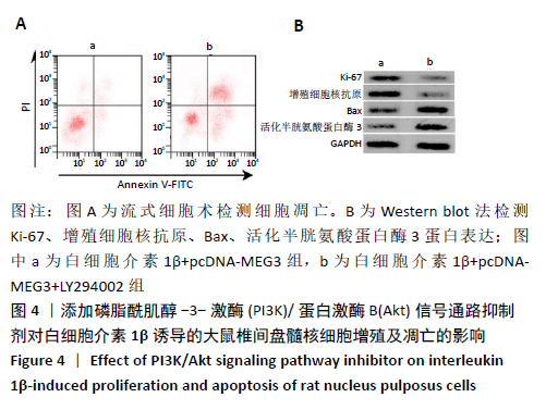
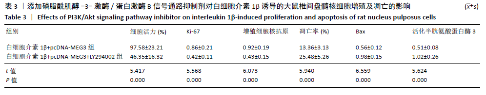
|