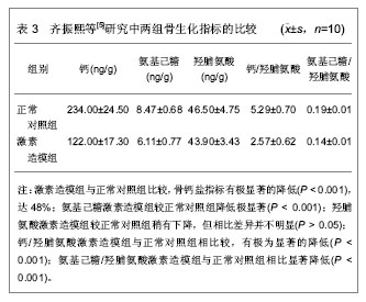
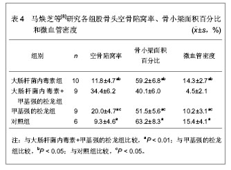
激素性股骨头坏死的发病机理
1.脂质代谢紊乱学说
正常情况下,肝内脂肪含量占肝总重量的5%,血中的游离脂肪酸被肝脏吸收后,在肝细胞内转化成甘油三酸酯与特殊蛋白结合以VLDL(very low density lipoprtein)形式释放入血液,经活性脂蛋白脂酶的作用后被组织利用,充分证明应用激素后,动物皮下脂肪分解成游离脂肪酸释放于血液,形成高脂血症,如果进入到肝细胞内的游离脂肪超过了肝脏将甘油三酸酯转化为VLDL的能力时,肝脏吸收游离脂肪酸转化的甘油三酸酯,必然将堆积在肝细胞内,最终形成脂肪肝,肝脏一方面释放出VLDL及乳糜微粒供周围组织利用,另一方面血内极低密度前β脂蛋白乳化不全,脂蛋白相互联合,在周围血流中构成脂肪栓子。
2.骨质疏松学说
糖皮质激素对整体骨骼产生双方面的影响,即一方面降低卵巢、睾丸、肾上腺的性激素合成与分泌,减少胃肠钙吸收,增加肾脏钙排泄,引起继发甲旁亢;另一方面, 长期应用皮质激素直接抑制成骨细胞的功能,刺激破骨细胞的活性,增加骨组织对PTH和1,25-(OH)2-D3的敏感性,在总体上造成成骨作用减弱,破骨作用加强。其结果骨小梁变细、疏松、萎缩或断裂,发生细微骨折,负重时,拱形结构在机械力的作用下,发生微细骨折而塌陷,继而进一步压迫骨内微血管引起缺血,最终导致股骨头坏死
3.血液高粘滞状态学说
大剂量应用糖皮质激素可以使血中纤维蛋白原升高,由于纤维蛋白原在血浆中形成网状结构,加之红细胞聚集,使血液粘度增加,而致微循环灌注量下降,此乃激素引起股骨头坏死的一个重要因素。
4.血管内凝血学说
早在1951年Cosgriff首先证明了激素能引起系统的高凝状态,他认为凝学异常是激素引起骨坏死的一个潜在因素。
4.1 SANFH具备血栓形成的条件 血栓形成需具备3个条件:血流缓慢;血液凝固性增高;血管内皮细胞受损。股骨头软骨下骨区域的显微结构使血流易于淤滞,此处终末动脉与迂曲拱形的终末毛细血管相连,尤其在局部血管收缩因子如内皮素等存在的情况下,有利于血栓形成。股骨头内小静脉被骨髓内肥大的脂肪细胞压迫也引起毛细血管骨血流淤积。血流淤积、血管内皮损伤和高凝三个因素造成循环内血栓形成。激素致骨内脂肪栓塞的机械堵塞和坏死部位局部的解剖特殊性使血流淤滞,游离脂肪酸的水解及其它因素如氧自由基损害血管内皮结构,骨内微循环(终末动脉、毛细血管、血窦)内皮细胞损害最有可能激活血小板聚集和纤维蛋白血栓形成,以后逐渐波及小静脉、静脉、小动脉和骨外动脉。激素所致血液高粘度、高凝、高脂血症和纤溶下降更具备了血栓形成的条件,加之缺血再灌注损伤和继发纤溶所致纤溶下降和局部内皮素和血栓素A2所致血管收缩,更容易在局部血栓形成。
4.2 股骨头血管内凝血导致股骨头坏死 股骨头内血栓形成,一方面,将损害动脉灌注,而且更大程度上亦损害静脉引流,后者造成骨内间室综合征,使骨内压上升,灌注下降,加重股骨头缺血以至坏死,即进行性缺血学说;另一方面,激活的凝血瀑布反应产生了炎症反应,进而加剧了局部损害,同时,继发纤溶使部分血栓溶解,尤其动脉内皮细胞膜脂质过氧化,致使骨髓内出血,进一步加重了股骨头的损害,导致股骨头的坏死。
4.3 SANFH股骨头血管内凝血的组织学研究 激素的直接细胞毒性作用造成血管损伤,导致动脉血供中断和骨髓内反复出血,从而引起股骨头坏死。以往由于骨内血栓染色技术的限制未能直接观察血栓的存在,使血管内凝血学说缺乏证据。
4.4 SANFH血管内凝血的血液学研究 血栓前状态(PTS)的存在已成为SANFH的一个不可忽视的因素。PTS是多种因素引起的止血、凝血和抗凝系统失调的一种病理过程,具有易导致血栓形成的多种血液学变化。内皮素是主要源于血管内皮细胞的一族血管活性物质,分为3型,以内皮素-1(ET-1)的活性最强。内皮素是迄今所知作用最强、持续时间最长的缩血管多肽,其血管收缩强度是血管紧张素的10倍,去甲肾上腺素的100倍。激素使机体内脂质代谢紊乱,产生高脂血症是造成血管内皮细胞损伤、内皮素分泌释放的主要原因。因为高脂血症时血中低密度脂蛋白和氧化型低密度脂蛋白增多与血管内皮细胞上的受体结合,通过三磷酸肌醇细胞内信息传递系统促进内皮素合成和释放;它们也可直接损伤血管内皮细胞使其内皮素释放增多;氧化型低密度脂蛋白还可刺激巨噬细胞合成和分泌内皮素。在生理情况下体内ET-1含量甚微,在病理或使用糖皮质激素情况下ET-1含量升高,可以使血管强烈收缩,且对静脉收缩明显强于对动脉的收缩作用。这样会在股骨头内形成高灌低排、髓内血液淤滞、骨内高压,且血液高凝,易于血栓形成。
5.静脉内瘀滞,引起的骨内高压学说
激素可使血小板聚集,血管闭塞,局部酸性代谢物质积聚,毛细血管通透性增高,血浆外渗,骨髓间质水肿,骨内压升高,导致股骨头坏死。此外,激素引起脂肪代谢紊乱,血脂升高,一方面骨髓内脂肪细胞堆积,压力增高,血液循环障碍,另一方面成骨障碍,共同导致股骨头坏死。
6.免疫复合物抗体沉积引起动脉血管炎 单纯激素仅能诱导出轻度骨内小动脉炎,而采用马血清造成家兔过敏性血管炎,再用激素可加重血清致敏后的血管炎。小动脉是血管炎的靶器官,免疫复合物沉积在血管壁能引起血管炎,而皮质类固醇能抑制胶原和弹性纤维的合成,对已有血管炎损害的血管可加重血管收缩,血小板聚集和内皮细胞增生,这些变化能引起小动脉断裂和栓塞,从而导致骨坏死。股骨头内小动脉为中末动脉,一旦损害则侧支循环难以代偿,由此造成的缺血易引起骨坏死。


.jpg)
.jpg)
.jpg)
.jpg)
.jpg)