中国组织工程研究 ›› 2013, Vol. 17 ›› Issue (10): 1809-1813.doi: 10.3969/j.issn.2095-4344.2013.10.016

• 干细胞培养与分化 stem cell culture and differentiation • 上一篇    下一篇

低氧环境下白细胞介素1β诱导中脑源性神经干细胞的分化

丁继固1,丁文杰2,李 光3   

  1. 1 湖北科技学院人体解剖学教研室,湖北省咸宁市 437100
    2 湖北科技学院口腔学教研室,湖北省咸宁市 437100
    3 武汉大学中南医院重症医学科,湖北省武汉市 43007
  • 收稿日期:2012-03-01 修回日期:2012-05-16 出版日期:2013-03-05 发布日期:2013-03-05
  • 作者简介:丁继固,男,1954年生,湖北省咸宁市人,汉族,1977年咸宁学院毕业,教授,硕士生导师,主要从事神经解剖学和应用解剖学方面的研究。 dingjigu@hotmail.com
  • 基金资助:

    湖北省教育厅重点课题(D20092802)。

Differentiation of mesencephalic neural stem cells induced by interleukin-1beta under hypoxic condition

Ding Ji-gu1, Ding Wen-jie2, Li Guang3   

  1. 1 Department of Human Anatomy, Hubei University of Science and Technology, Xianning 437100, Hubei Province, China
    2 Department of Stomatology, Hubei University of Science and Technology, Xianning 437100, Hubei Province, China
    3 Department of Critical Care Medicine, Wuhan 430071, Hubei Province, China
  • Received:2012-03-01 Revised:2012-05-16 Online:2013-03-05 Published:2013-03-05
  • About author:Ding Ji-gu, Professor, Master’s supervisor, Department of Human Anatomy, Hubei University of Science and Technology, Xianning 437100, Hubei Province, China dingjigu@hotmail.com

摘要:

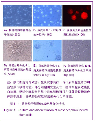
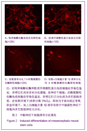
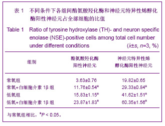
背景:在体外某些外来信号的调控和诱导下,神经干细胞可定向诱导分化成与受体部位细胞类型与构成相似的细胞群体,使其更容易掺入靶组织,并可满足移植所需的细胞数量,提高移植细胞的存活率。
目的:验证白细胞介素1β在低氧环境下体外诱导中脑源性神经干细胞的分化。
方法:分离孕12 d胎鼠中脑组织,培养神经干细胞、传代并鉴定。将鉴定后传代培养的中脑源性神经干细胞按分组接种于含体积分数10%胎牛血清的DMEM/F12培养基和含体积分数10%胎牛血清的DMEM/ F12+白细胞介素1β培养基;分别置于常氧(体积分数21%O2)和低氧(体积分数3%O2)环境下诱导分化,诱导分化9-11 d后行小鼠抗大鼠酪氨酸羟化酶免疫细胞化学染色,检测酪氨酸羟化酶及神经元特异性烯醇化酶阳性细胞率。
结果与结论:与常氧组比较,低氧组尤其是低氧+白细胞介素1β组中酪氨酸羟化酶阳性神经元的突起数目更多,且长度更为延长。孕12 d胚鼠腹侧中脑组织可以在体外培养传代、并能诱导分化成多巴胺能神经元;在低氧环境或低氧+白细胞介素1β诱导下,分化率均高于常氧组,其表型更成熟。说明在低氧或低氧和白细胞介素1β诱导下,可明显促进中脑源性干细胞分化为形态及功能成熟的多巴胺能神经元。

关键词: 干细胞, 干细胞培养与分化, 低氧环境, 白细胞介素1β, 中脑源性干细胞, 酪氨酸羟化酶, 多巴胺能神经元, 省级基金, 干细胞图片文章

Abstract:

BACKGROUND: Under the regulation and induction by some in vitro signals, neural stem cells can be induced to a cell population with similar types and composition to the cells in the receptor region, which facilitates cells to easily incorporate into target tissue, meets the cell quantity required by cell transplantation and increases the survival rate of transplanted cells.
OBJECTIVE: To validate the differentiation of mesencephalic neural stem cells induced by interleukin 1β under hypoxic conditions.
METHODS: Following harvesting mesencephalic tissue from 12 day pregnant fetal mice, neural stem cells were cultured and identified. Then these mesencephalic neural stem cells were inoculated into DMEM/F12 culture medium containing 10% fetal bovine serum and DMEM/F12 culture medium containing 10% fetal bovine serum + interleukin 1β under normoxic (21%O2) and hypoxic (3% O2) environment. After 9-11 days of induced differentiation, immunohistochemical staining and flow cytometry were performed to detect tyrosine hydroxylase- and neuronspecific enolase-positive expression. Neural stem cells of mesencephalic tissue of 12-day pregnant mice can be in vitro cultured and induce-differentiated into dopaminergic neurons. Under hypoxic environment or hypoxic environment + interleukin 1β induction, mesencephalic neural stem cells showed a high differentiation rate and more matured phenotype compared to the normoxic conditions. These findings suggest that hypoxia or hypoxia + interleukin 1β can significantly promote mesencephalic neural stem cells to differentiate into dopaminergic neurons with more matured morphology and functions.

Key words: stem cells, stem cell culture and differentiation, hypoxia, interleukin-1β, mesencephalic neural stem cell, tyrosine hydroxylase, dopaminergic neuron, provincial grants-supported paper, stem cell photographs-containing paper

中图分类号: