| [1] McKay R.Stem cells in the central nervous system.Science. 1997;276(5309):66-71.
[2] Reynolds BA, Tetzlaff W, Weiss S.A multipotent EGF-responsive striatal embryonic progenitor cell produces neurons and astrocytes.J Neurosci. 1992;12(11):4565-4574.
[3] Arvidsson A, Collin T, Kirik D,et al. Neuronal replacement from endogenous precursors in the adult brain after stroke. Nat Med. 2002;8(9):963-970.
[4] 韩宁.白藜芦醇保护神经干细胞氧糖剥夺损伤的实验研究[D].西安:第四军医大学,2013.
[5] 包志军.川芎嗪对谷氨酸损伤海马神经元保护作用的研究[D].苏州:苏州大学,2009.
[6] 李国栋,鄢文海,邢莹.五味子酮对APP转基因鼠神经干细胞分化中Tau蛋白过磷酸化水平的影响[J].中国组织工程研究与临床康复,2009,13(23):4490-4494.
[7] 国家药典委员会.中华人民共和国药典[M].北京:化学工业出版社, 2010.
[8] 周爱珠.平衡罐合独活寄生汤治疗功能性背腰痛[J].中医外治杂志,2014,23(2):20-21.
[9] 东方闻睿.神农本草经[M].北京:中国电影出版社,2001.
[10] Okuyama T, Takata M, Nishino H,et al.Studies on the antitumor-promoting activity of naturally occurring substances. II. Inhibition of tumor-promoter-enhanced phospholipid metabolism by umbelliferous materials.Chem Pharm Bull (Tokyo). 1990;38(4):1084-1086.
[11] 胡昱,赵丹,张晓丹,等.独活不同提取部位抑制H2O2诱导的SH-SY5Y细胞损伤[J].中国实验方剂学杂志,2013,19(24): 184-188.
[12] 裴媛.独活及其醇提物延缓自然衰老小鼠脑老化的实验研究[D].沈阳:辽宁中医学院,2005.
[13] Yang J, Jiang Z, Fitzgerald DC,et al.Adult neural stem cells expressing IL-10 confer potent immunomodulation and remyelination in experimental autoimmune encephalitis.J Clin Invest. 2009;119(12):3678-3691.
[14] Yang J, Bridges K, Chen KY,et al.Riluzole increases the amount of latent HSF1 for an amplified heat shock response and cytoprotection.PLoS One. 2008;3(8):e2864.
[15] Escribano L, Simón AM, Gimeno E,et al.Rosiglitazone rescues memory impairment in Alzheimer's transgenic mice: mechanisms involving a reduced amyloid and tau pathology. Neuropsychopharmacology. 2010;35(7): 1593-1604.
[16] Shi Y, Sun G, Zhao C,et al.Neural stem cell self-renewal.Crit Rev Oncol Hematol. 2008;65(1):43-53.
[17] Nyfeler Y, Kirch RD, Mantei N,et al.Jagged1 signals in the postnatal subventricular zone are required for neural stem cell self-renewal.EMBO J. 2005;24(19):3504-3515.
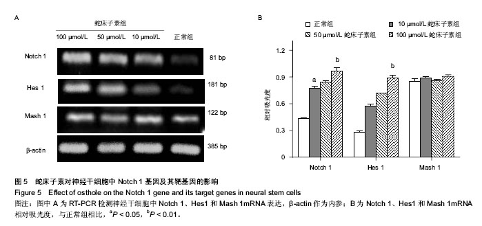
[18] 刘菲菲,徐丽萍,龙大宏,等.神经干细胞的单层培养及分化过程中HES1和MASH1的表达变化[J].中山大学学报:医学科学版, 2014,35(1):18-24.
[19] 尚红记,刘运林.Notch信号通路参与外源性碱性成纤维细胞生长因子对神经干细胞辐射损伤保护作用的实验研究[J].中华行为医学与脑科学杂志,2014,23(1):12-14.
[20] 龙海波.大鼠损伤脊髓提取液对胚胎大鼠神经干细胞增殖能力及 Notch1、Hes1表达的影响[D]. 泸州:泸州医学院,2013.
[21] Piccin D, Yu F, Morshead CM.Notch signaling imparts and preserves neural stem characteristics in the adult brain.Stem Cells Dev. 2013;22(10):1541-1550.
[22] Dong Z, Yang N, Yeo SY,et al.Intralineage directional Notch signaling regulates self-renewal and differentiation of asymmetrically dividing radial glia.Neuron. 2012;74(1):65-78.
[23] Nakamura Y, Sakakibara Si, Miyata T,et al.The bHLH gene hes1 as a repressor of the neuronal commitment of CNS stem cells.J Neurosci. 2000;20(1):283-293. |