| [1]Puche JE, Saiman Y, Friedman SL.Hepatic stellate cells and liver fibrosis.Compr Physiol. 2013;3(4):1473-1492.
[2]胡昆鹏,林楠,林继宗,等.人骨髓间质干细胞对肝星状细胞的体外调控[J].中国组织工程研究与临床康复,2009,13(27): 5257-5260.
[3]曹葆强,林继宗,钟跃思,等.自体骨髓细胞经门静脉移植治疗肝硬化与肝功能不全的临床研究[J].中华普通外科杂志,2007,22(5): 386-389.
[4]Lin N, Tang Z, Deng M,et al.Hedgehog-mediated paracrine interaction between hepatic stellate cells and marrow-derived mesenchymal stem cells.Biochem Biophys Res Commun. 2008;372(1):260-265.
[5]钟跃思,林楠,潘楚芝,等.氯甲基苯甲酰氨荧光染料标记大鼠骨髓间质干细胞的体内示踪[J].中国组织工程研究与临床康复,2008, 12(51):10090-10094.
[6]Friedman SL.Hepatic stellate cells: protean, multifunctional, and enigmatic cells of the liver.Physiol Rev. 2008;88(1):125-172.
[7]Roderfeld M, Weiskirchen R, Wagner S,et al.Inhibition of hepatic fibrogenesis by matrix metalloproteinase-9 mutants in mice.FASEB J. 2006;20(3):444-454.
[8]Baba S, Fujii H, Hirose T,et al.Commitment of bone marrow cells to hepatic stellate cells in mouse.J Hepatol. 2004;40(2): 255-260.
[9]Russo FP, Alison MR, Bigger BW,et al.The bone marrow functionally contributes to liver fibrosis.Gastroenterology. 2006;130(6):1807-1821.
[10]De Minicis S, Seki E, Uchinami H,et al.Gene expression profiles during hepatic stellate cell activation in culture and in vivo.Gastroenterology. 2007;132(5):1937-1946.
[11]Lin N, Hu K, Chen S,et al.Nerve growth factor-mediated paracrine regulation of hepatic stellate cells by multipotent mesenchymal stromal cells.Life Sci. 2009;85(7-8):291-295.
[12]杨文,覃山羽,姜海行,等.骨髓间充质干细胞体外调控肝星状细胞死亡受体5的表达[J]. 中国组织工程研究,2012,16(27): 4947-4952.
[13]Sasore T, Reynolds AL, Kennedy BN.Targeting the PI3K/Akt/mTOR Pathway in Ocular Neovascularization.Adv Exp Med Biol. 2014;801:805-811.
[14]Magi S, Saeki Y, Kasamatsu M,et al.Chemical genomic-based pathway analyses for epidermal growth factor-mediated signaling in migrating cancer cells.PLoS One. 2014;9(5): e96776.
[15]Jung KH, Yan HH, Fang Z,et al.HS-104, a PI3K inhibitor, enhances the anticancer efficacy of gemcitabine in pancreatic cancer.Int J Oncol. 2014;45(1):311-321.
[16]Martelli AM, Lonetti A, Buontempo F, et al.Targeting Signaling Pathways in T-cell acute lymphoblastic leukemia initiating cells.Adv Biol Regul. 2014. [Epub ahead of print]
[17]Nair S, Hagberg H, Krishnamurthy R,et al.Death associated protein kinases: molecular structure and brain injury.Int J Mol Sci. 2013;14(7):13858-13872.
[18]Puustinen P, Rytter A, Mortensen M,et al.CIP2A oncoprotein controls cell growth and autophagy through mTORC1 activation.J Cell Biol. 2014;204(5):713-727.
[19]Fallet V, Cadranel J, Doubre H,et al.Prospective screening for ALK: clinical features and outcome according to ALK status. Eur J Cancer. 2014;50(7):1239-1246.
[20]Rafiq K, Kolpakov MA, Seqqat R,et al. c-Cbl Inhibition Improves Cardiac function and Survival in Response to Myocardial Ischemia.Circulation. 2014. [Epub ahead of print]
[21]Zerenturk EJ, Sharpe LJ, Ikonen E,et al.Desmosterol and DHCR24: unexpected new directions for a terminal step in cholesterol synthesis.Prog Lipid Res. 2013;52(4):666-680.
[22]Fehér A, Juhász A, Pákáski M,et al.Gender dependent effect of DHCR24 polymorphism on the risk for Alzheimer's disease. Neurosci Lett. 2012;526(1):20-23.
[23]Zerenturk EJ, Sharpe LJ, Brown AJ.Sterols regulate 3β-hydroxysterol Δ24-reductase (DHCR24) via dual sterol regulatory elements: cooperative induction of key enzymes in lipid synthesis by Sterol Regulatory Element Binding Proteins.Biochim Biophys Acta. 2012;1821(10):1350-1360.
[24]Sarajärvi T, Lipsanen A, Mäkinen P,et al.Bepridil decreases Aβ and calcium levels in the thalamus after middle cerebral artery occlusion in rats.J Cell Mol Med. 2012;16(11):2754- 2767.
[25]Halepoto DM, Bashir S, A L-Ayadhi L.Possible role of brain-derived neurotrophic factor (BDNF) in autism spectrum disorder: current status.J Coll Physicians Surg Pak. 2014; 24(4):274-278.
[26]Keleshian VL, Kellom M, Kim HW,et al.Neuropathological Responses to Chronic NMDA in Rats Are Worsened by Dietary n-3 PUFA Deprivation but Are Not Ameliorated by Fish Oil Supplementation.PLoS One. 2014;9(5):e95318.
[27]Pla P, Orvoen S, Saudou F, et al.Mood disorders in Huntington's disease: from behavior to cellular and molecular mechanisms.Front Behav Neurosci. 2014;8:135.
[28]Takei N, Nawa H.mTOR signaling and its roles in normal and abnormal brain development.Front Mol Neurosci. 2014;7:28.
[29]Azizidoost S, Shanaki Bavarsad M, Shanaki Bavarsad M, et al. The role of notch signaling in bone marrow niche. Hematology. 2014. [Epub ahead of print]
[30]Suila H, Hirvonen T, Ritamo I,et al.Extracellular o-linked N-acetylglucosamine is enriched in stem cells derived from human umbilical cord blood.Biores Open Access. 2014; 3(2):39-44.
[31]Kececi Y, Sir E.Prediction of resection weight in reduction mammaplasty based on anthropometric measurements. Breast Care (Basel). 2014;9(1):41-45.
[32]Li X, Martinez-Fernandez A, Hartjes KA,et al.Transcriptional Atlas of Cardiogenesis Maps Congenital Heart Disease Interactome.Physiol Genomics. 2014. [Epub ahead of print] |

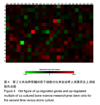
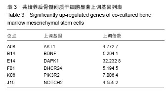
.jpg)