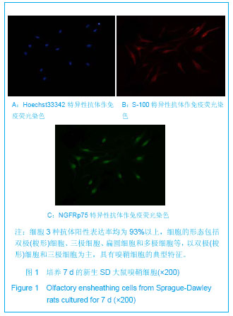
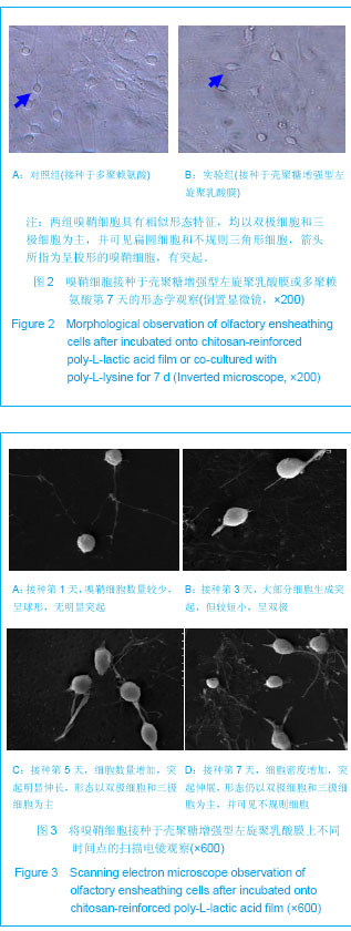
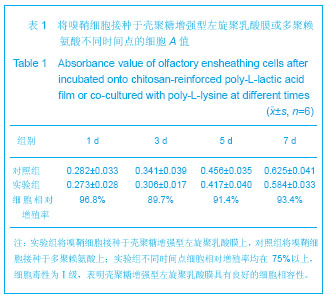
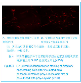
| [1] Potter W,Kalil RE,Kao WJ. Biomimetic material systems for neural progenitor cell-based therapy. Front Biosci.2008;13: 806-821.[2] Chen BK,Knight AM,de Ruiter GC,et al.Axon regeneration through scaffold into distal spinal cord after transection.J Neurotrauma.2009;26(10):1759-1771.[3] Kubinová S,Syková E.Biomaterials combined with cell therapy for treatment of spinal cord injury.Regen Med.2012; 7(2):207-224.[4] Cheng H,Huang YC,Chang PT,et al.Laminin-incorporated nerve conduits made by plasma treatment for repairing spinal cord injury. Biochem Biophys Res Commun. 2007;357(4): 938-944.[5] Li XG,Yang ZY,Zhang AF,et al.Repair of thoracic spinal cord injury by chitosan tube implantationin adult rats.Biomaterials. 2009;30(6):1121-1132.[6] Giannetti S,Lauretti L,Fernandez E,et al.Acrylic hydrogel implants after spinal cord lesion in the adult rat.Neural Res. 2001;23(4):405-409.[7] Hurtado A,Moon LDF,Maquet V,et al.Poly (D,L-lactic acid) macroporous guidance scaffolds seeded with Schwann cells genetically modified to secrete a bi-functional neurotrophin implanted in the completely transected adult rat thoracic spinal cord. Biomaterials.2006;27(3):430-442.[8] Moore MJ,Friedman JA,Lewellyn EB,et al.Multiple-channel scaffolds to promote spinal cord axon regeneration. Biomaterials. 2006;27(3):419-429.[9] Xu XM,Zhang SX,Li H,et al.Regrowth of axons into the distal spinal cord through a Schwann-cell-seeded mini-channel implanted into hemisected adult rat spinal cord.Eur J Neurosci.1999;11(5):1723-1740.[10] Woerly S,Pinet E,Robertis L,et al.Spinal cord repair with PHPMA hydrogel containing RGD peptides(NeuroGelTM). Biomaterials.2001;22(10):1095-1111.[11] Woerly S,Doan VD,Sosa N,et al.Prevention of gliotic scar formation by neurogel allows partial endogenous repair of transected cat spinal cord. J Neurosci Res.2004;75(2): 262-272.[12] Tharion G,Indirani K,Durai M,et al.Motor recovery following olfactory ensheathing cell transplantation in rats with spinal cord injury. Neurol India.2011;59(4):566-572.[13] Lopes A.Olfactory ensheathing cells for human spinal cord injury. Neurorehabil Neural Repair.2010;24(8):772-773.[14] Cui YL,Qi AD,Liu WG,et al. Biomimetic surface modification of poly(L-lactic acid) with chitosan and its effects on articular chondrocytes in vitro.Biomaterials.2003;24(21):3859-3868.[15] Novikova LN,Lobov S,Wiberg M,et al. Efficacy of olfactory ensheathing cells to support regeneration after spinal cord injury is influenced by method of culture preparation.Exp Neurol.2011;229(1):132-142.[16] Harrop JS,Hashimoto R,Norvell D,et al.Evaluation of clinical experience using cell-based therapies in patients with spinal cord injury: a systematic review.J Neurosurg Spine.2012; 17(1):230-246.[17] Ozdemir M,Attar A,Kuzu I. Regenerative treatment in spinal cord injury. Curr Stem Cell Res Ther.2012;7(5):364-369.[18] Yazdani SO,Pedram M,Hafizi M,et al. A comparison between neurally induced bone marrow derived mesenchymal stem cells and olfactory ensheathing glial cells to repair spinal cord injuries in rat. Tissue Cell.2012;44(4):205-213.[19] Nash HH,Borke RC,Anders JJ. New method of purification for establishing primary cultures of ensheathing cells from the adult olfactory bulb.Glia.2001;34(2):81-87.[20] Jani HR,Raisman G.Ensheathing cell cultures from the olfactory bulb and mucosa.Glia.2004;47(2):130-137.[21] Crapo PM, Medberry CJ, Reing JE,et al. Biologic scaffolds composed of central nervous system extracellular matrix. Biomaterials.2012;33(13):3539-3547.[22] Novikova LN, Novikov LN, Kellerth JO. Biopolymers and biodegradable smart implants for tissue regeneration after spinal cord injury. Curr Opin Neurol.2003;16(6):711-715.[23] Novikov LN, Novikova LN, Mosahebi A,et al. A novel biodegradable implant for neuronal rescue and regeneration after spinal cord injury. Biomaterials.2002;23(16):3369-3376.[24] Jin L, Feng ZQ, Zhu ML,et al. A novel fluffy conductive polypyrrole nano-layer coated PLLA fibrous scaffold for nerve tissue engineering. J Biomed Nanotechnol. 2012;8(5): 779-785.[25] Boccafoschi F, Fusaro L, Mosca C,et al. The biological response of poly(L-lactide) films modified by different biomolecules: role of the coating strategy. J Biomed Mater Res A.2012;100(9):2373-2381.[26] Huang WC, Yao CK, Liao JD,et al. Enhanced Schwann cell adhesion and elongation on a topographically and chemically modified poly(L-lactic acid) film surface. J Biomed Mater Res A.2011;99(2):158-165.[27] Zheng L, Cui HF. Enhancement of nerve regeneration along a chitosan conduit combined with bone marrow mesenchymal stem cells. J Mater Sci Mater Med. 2012;23(9):2291-2302.[28] Yi X, Jin G, Tian M,et al. Porous chitosan scaffold and NGF promote neuronal differentiation of neural stem cells in vitro. Neuro Endocrinol Lett. 2011;32(5):705-710.[29] Li X, Liu X, Dong W,et al. In vitro evaluation of porous poly(L-lactic acid) scaffold reinforced by chitin fibers.J Biomed Mater Res B Appl Biomater.2009;90(2):503-509.[30] Zhao M, Li L, Li X, et al. Three-dimensional honeycomb-patterned chitosan/poly(L-lactic acid) scaffolds with improved mechanical and cell compatibility.J Biomed Mater Res A. 2011;98(3):434-441.[31] Ku Y, Shim IK, Lee JY, et al. Chitosan/poly(L-lactic acid) multilayered membrane for guided tissue regeneration.J Biomed Mater Res A.2009;90(3):766-772.[32] Jiao Y, Liu Z, Zhou C. Fabrication and characterization of PLLA-chitosan hybrid scaffolds with improved cell compatibility.J Biomed Mater Res A.2007;80(4):820-825.[33] Liu Z, Jiao Y, Zhang Z, et al. Surface modification of poly(L-lactic acid) by entrapment of chitosan and its derivatives to promote osteoblasts-like compatibility.J Biomed Mater Res A.2007;83(4):1110-1116.[34] 徐冠华,崔志明,黄镇,等.纤维蛋白凝胶与大鼠嗅鞘细胞的生物相容性[J].中国组织工程研究与临床康复,2009,13(47): 9249-9252.[35] Nojehdehian H, Moztarzadeh F, Baharvand H,et al. Effect of poly-L-lysine coating on retinoic acid-loaded PLGA microspheres in the differentiation of carcinoma stem cells into neural cells.Int J Artif Organs.2010;33(10):721-730.[36] Martín-López E, Nieto-Díaz M, Nieto-Sampedro M. Differential adhesiveness and neurite-promoting activity for neural cells of chitosan, gelatin, and poly-L-lysine films. J Biomater Appl.2012;26(7):791-809. |
.jpg)