[1] GADALETA RM, MOSCHETTA A. Metabolic Messengers: fibroblast growth factor 15/19. Nat Metab. 2019;1(6):588-594.
[2] FREIIN VON HÖVEL F, KEFALAKES E, GROTHE C. What Can We Learn from FGF-2 Isoform-Specific Mouse Mutants? Differential Insights into FGF-2 Physiology In Vivo. Int J Mol Sci. 2020;22(1):390.
[3] JIA T, JACQUET T, DALONNEAU F, et al. FGF-2 promotes angiogenesis through a SRSF1/SRSF3/SRPK1-dependent axis that controls VEGFR1 splicing in endothelial cells. BMC Biol. 2021;19(1):173.
[4] NOVAIS A, CHATZOPOULOU E, CHAUSSAIN C, et al. The Potential of FGF-2 in Craniofacial Bone Tissue Engineering: A Review. Cells. 2021;10(4):932.
[5] OTSUKA T, MENGSTEAB PY, LAURENCIN CT. Control of mesenchymal cell fate via application of FGF-8b in vitro. Stem Cell Res. 2021;51:102155.
[6] CHEN K, RAO Z, DONG S, et al. Roles of the fibroblast growth factor signal transduction system in tissue injury repair. Burns Trauma. 2022;10:tkac005.
[7] HOSAKA K, YANG Y, SEKI T, et al. Therapeutic paradigm of dual targeting VEGF and PDGF for effectively treating FGF-2 off-target tumors. Nat Commun. 2020;11(1):3704.
[8] PAJAZITI B, YOSY K, STEINBERG OV, et al. FGF-23 protects cell function and viability in murine pancreatic islets challenged by glucolipotoxicity. Pflugers Arch. 2023;475(3):309-322.
[9] KATOH M. FGFR inhibitors: Effects on cancer cells, tumor microenvironment and whole-body homeostasis (Review). Int J Mol Med. 2016;38(1):3-15.
[10] FRANCAVILLA C, O’BRIEN CS. Fibroblast growth factor receptor signalling dysregulation and targeting in breast cancer. Open Biol. 2022;12(2):210373.
[11] LI X. The FGF metabolic axis. Front Med. 2019;13(5):511-530.
[12] KUMAR V, GOUTAM RS, PARK S, et al. Functional Roles of FGF Signaling in Early Development of Vertebrate Embryos. Cells. 2021;10(8):2148.
[13] 欧明明,黄晓峰,韩培彦.成纤维细胞生长因子与器官发育[J].中国组织工程研究与临床康复,2011,15(15):2800-2804.
[14] ARDIZZONE A, SCUDERI SA, GIUFFRIDA D, et al. Role of Fibroblast Growth Factors Receptors (FGFRs) in Brain Tumors, Focus on Astrocytoma and Glioblastoma. Cancers (Basel). 2020;12(12):3825.
[15] EBEID M, HUH SH. FGF signaling: diverse roles during cochlear development. BMB Rep. 2017;50(10):487-495.
[16] FAROOQ M, KHAN AW, KIM MS, et al. The Role of Fibroblast Growth Factor (FGF) Signaling in Tissue Repair and Regeneration. Cells. 2021;10(11):3242.
[17] KATOH M. Fibroblast growth factor receptors as treatment targets in clinical oncology. Nat Rev Clin Oncol. 2019;16(2):105-122.
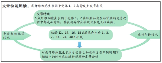
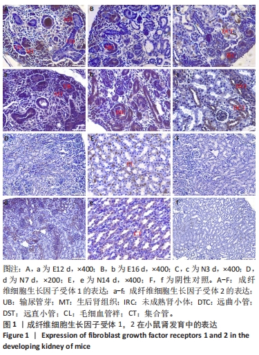
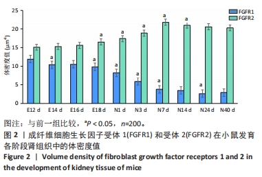
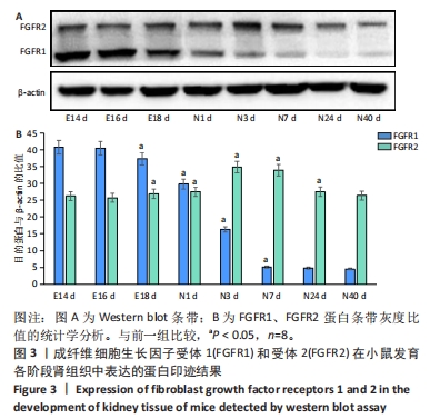
[18] 田娟,郭敏,王堃.成纤维细胞生长因子受体1和受体2在小鼠肾发育中的表达[J].解放军医学杂志,2010,35(12):1471-1474.
[19] XIE Y, SU N, YANG J, et al. FGF/FGFR signaling in health and disease. Signal Transduct Target Ther. 2020;5(1):181.
[20] 薄双玲,马太芳,田鹤,等.小鼠肾小管组织中水通道蛋白1、7的动态表达和意义[J].遵义医科大学学报,2021,44(2):179-182,187.
[21] 宋科昕.发生发育小鼠肾小球形态计量学研究[D].沈阳:中国医科大学,2020.
[22] BREWER JR, MAZOT P, SORIANO P. Genetic insights into the mechanisms of Fgf signaling. Genes Dev. 2016;30(7):751-771.
[23] ROSKOSKI R JR. The role of fibroblast growth factor receptor (FGFR) protein-tyrosine kinase inhibitors in the treatment of cancers including those of the urinary bladder. Pharmacol Res. 2020;151:104567.
[24] TRUEB B, AMANN R, GERBER SD. Role of FGFRL1 and other FGF signaling proteins in early kidney development. Cell Mol Life Sci. 2013;70(14):2505-2518.
[25] BATES CM. Role of fibroblast growth factor receptor signaling in kidney development. Pediatr Nephrol. 2011;26(9):1373-1379.
[26] QIAO J, BUSH KT, STEER DL, et al. Multiple fibroblast growth factors support growth of the ureteric bud but have different effects on branching morphogenesis. Mech Dev. 2001;109(2):123-135.
[27] WALKER KA, SIMS-LUCAS S, BATES CM. Fibroblast growth factor receptor signaling in kidney and lower urinary tract development. Pediatr Nephrol. 2016;31(6):885-895.
[28] ZHAO H, KEGG H, GRADY S, et al. Role of fibroblast growth factor receptors 1 and 2 in the ureteric bud. Dev Biol. 2004;276(2):403-415.
[29] CHAN K, LI X. Current Epigenetic Insights in Kidney Development. Genes (Basel). 2021; 12(8):1281.
[30] 徐卓.成纤维细胞生长因子家族在肾脏疾病中的作用和机制研究[D].南京:南京医科大学,2014. |