中国组织工程研究 ›› 2024, Vol. 28 ›› Issue (20): 3190-3195.doi: 10.12307/2024.333

• 骨组织构建 bone tissue construction • 上一篇    下一篇

miR-155/瘦素受体/AMPK轴在结核菌素诱导破骨细胞形成中的作用及机制

王增顺,索南昂秀,刘立民,周京元   

  1. 青海省人民医院骨科,青海省西宁市  810000
  • 收稿日期:2020-10-21 接受日期:2020-12-18 出版日期:2024-07-18 发布日期:2023-09-11
  • 通讯作者: 索南昂秀,主任医师,青海省人民医院骨科,青海省西宁市 810000
  • 作者简介:王增顺,男,1987年生,青海省海东市人,汉族,2013年大连医科大学毕业,硕士,主治医师,主要从事脊柱外科研究。
  • 基金资助:
    2017年青海省卫生和计划生育委员会科研课题(2017-wjzdx-05),项目负责人:索南昂秀

Role and mechanism of miR-155/leptin receptor/adenosine phosphate-dependent protein kinase axis in tuberculin-induced osteoclast formation

Wang Zengshun, Suonan Angxiu, Liu Limin, Zhou Jingyuan   

  1. Department of Orthopedics, Qinghai Provincial People’s Hospital, Xining 810000, Qinghai Province, China
  • Received:2020-10-21 Accepted:2020-12-18 Online:2024-07-18 Published:2023-09-11
  • Contact: Suonan Angxiu, Chief physician, Department of Orthopedics, Qinghai Provincial People’s Hospital, Xining 810000, Qinghai Province, China
  • About author:Wang Zengshun, Master, Attending physician, Department of Orthopedics, Qinghai Provincial People’s Hospital, Xining 810000, Qinghai Province, China
  • Supported by:
    2017 Scientific Research Project of Qinghai Provincial Health and Family Planning Commission, No. 2017-wjzdx-05 (to SNAX)

摘要:


文题释义:

miRNA:一族18-25 nt的小分子非编码RNA,能够识别靶基因mRNA 3’UTR并阻碍mRNA翻译或诱导mRNA水解,进而在转录后水平实现对基因表达的负调控。
破骨细胞:由单核巨噬细胞分化而来的终末细胞,介导骨吸收,在骨发育、生长、修复、重建中具有重要的作用。


背景:破骨细胞异常活化在脊柱结核骨质破坏中起重要作用。在骨质疏松发病过程中,敲低miR-155通过增加瘦素受体的表达来激活磷酸腺苷依赖的蛋白激酶(AMP-dependent protein kinase,AMPK),进而抑制破骨细胞分化及骨吸收。但miR-155/瘦素受体/AMPK轴在脊柱结核骨质破坏中的作用尚不清楚。

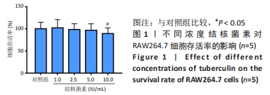
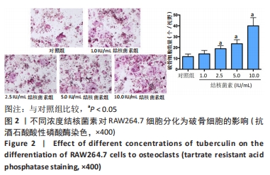
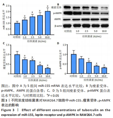
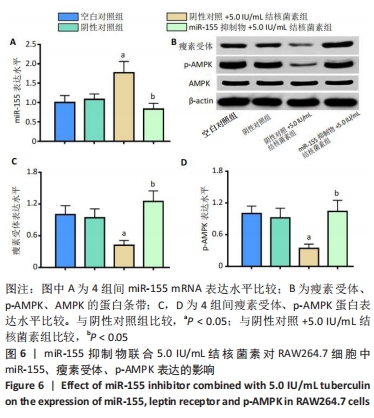
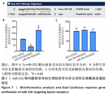
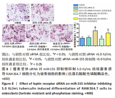
目的:研究miR-155/瘦素受体/AMPK轴在结核菌素诱导破骨细胞形成中的作用及机制。
方法:培养单核巨噬细胞RAW264.7细胞,用不同浓度(1.0,2.5,5.0,10.0 IU/mL)结核菌素处理,转染阴性对照序列或miR-155抑制物、阴性对照siRNA序列或瘦素受体siRNA序列。采用抗酒石酸酸性磷酸酶染色检测破骨细胞数量,荧光定量PCR检测miR-155 mRNA表达,Western blot检测瘦素受体、p-AMPK蛋白表达,双荧光素酶报告基因验证miR-155靶向瘦素受体。

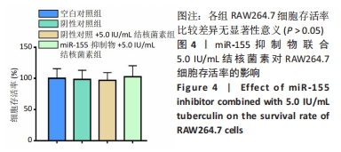
结果与结论:①与对照组比较,2.5,5.0,10.0 IU/mL结核菌素组破骨细胞数量、miR-155 mRNA表达水平明显增加,瘦素受体、p-AMPK蛋白表达水平明显降低(P < 0.05);②与阴性对照+5.0 IU/mL结核菌素组比较,miR-155抑制物+5.0 IU/mL结核菌素组破骨细胞数量、miR-155 mRNA表达水平明显降低,瘦素受体、p-AMPK蛋白表达水平明显增加(P < 0.05);③与阴性对照组比较,miR-155抑制物组瘦素受体野生型双荧光素酶报告基因的荧光活力增加,miR-155模拟物组瘦素受体野生型双荧光素酶报告基因的荧光活力降低(P < 0.05);④与阴性对照siRNA+miR-155抑制物+5.0 IU/mL结核菌素组比较,瘦素受体siRNA+miR-155抑制物+5.0 IU/mL结核菌素组miR-155 mRNA表达水平无明显变化(P > 0.05),破骨细胞数量明显增加,瘦素受体、p-AMPK蛋白表达水平明显降低(P < 0.05);⑤结果表明,结核菌素通过增加miR-155表达抑制下游瘦素受体表达及AMPK激活,进而诱导破骨细胞形成。

https://orcid.org/0000-0001-5959-2918(王增顺)

中国组织工程研究杂志出版内容重点:组织构建;骨细胞;软骨细胞;细胞培养;成纤维细胞;血管内皮细胞;骨质疏松;组织工程

关键词: 破骨细胞, 结核菌素, 单核巨噬细胞, 骨吸收, miR-155, 瘦素受体, AMPK

Abstract: BACKGROUND: Abnormal activation of osteoclasts plays an important role in the bone destruction due to spinal tuberculosis. During the pathogenesis of osteoporosis, miR-155 knockdown activates adenosine phosphate-dependent protein kinase (AMPK) by increasing the expression of leptin receptors, thereby inhibiting osteoclast differentiation and bone resorption. However, the role of miR-155/leptin receptor(LEPR)/AMPK axis in the bone destruction due to spinal tuberculosis remains unclear.
OBJECTIVE: To investigate the role and mechanism of miR-155/LEPR/AMPK axis in tuberculin-induced osteoclast formation. 
METHODS: RAW264.7 cells were cultured and treated with different concentrations of purified protein derivative (PPD) (1.0, 2.5, 5.0, 10.0 IU/mL) and transfected with negative control (NC) sequence or miR-155 inhibitor, NC siRNA sequence or LEPR siRNA sequence. Tartrate resistant acid phosphatase staining was used to detect the number of osteoclasts. Fluorescence quantitative PCR was used to detect the expression of miR-155. Western blot was used to detect the expression of LEPR and p-AMPK. Double luciferase reporter gene was used to verify miR-155 targeting LEPR. 
RESULTS AND CONCLUSION: Compared with the control group, the number of osteoclasts and the expression level of miR-155 significantly increased, while the expression level of LEPR and p-AMPK significantly decreased in 2.5, 5.0, and 10.0 IU/mL PPD groups (P < 0.05). Compared with NC+5.0 IU/mL PPD group, the number of osteoclasts and the expression level of miR-155 significantly decreased, while the expression level of LEPR and p-AMPK significantly increased in the miR-155 inhibitor+5.0 IU/mL PPD group (P < 0.05). Compared with the NC group, the fluorescence activity of LEPR wild-type double luciferase reporter gene was increased in the miR-155 inhibitor group, and decreased in the miR-155 mimic group (P < 0.05). Compared with si-NC+miR-155 inhibitor+5.0 IU/mL PPD group, the expression level of miR-155 had no significant change, the number of osteoclasts significantly increased, and the expression levels of LEPR and p-AMPL significantly decreased in si-LEPR+miR-155 inhibitor+5.0 IU/mL PPD group (P < 0.05). To conclude, tuberculin can induce osteoclast formation by increasing miR-155 expression and inhibiting downstream LEPR expression and AMPK activation.

Key words: osteoclast, tuberculin, mononuclear macrophages, bone resorption, miR-155, leptin receptor, AMPK

中图分类号: