中国组织工程研究 ›› 2023, Vol. 27 ›› Issue (36): 5864-5869.doi: 10.12307/2023.760

• 骨科植入物相关临床实践 Clinical practice of orthopedic implant • 上一篇    下一篇

动态对比增强核磁评价股骨头坏死微循环灌注的改变

环大维1,袁兆丰1,窦维强2,夏天卫1,邱  越1,张  超1,沈计荣1   

  1. 1南京中医药大学附属医院,江苏省中医院骨伤科,江苏省南京市   210000;2通用电气医疗集团磁共振研究部,北京市   100000
  • 收稿日期:2022-09-24 接受日期:2022-12-09 出版日期:2023-12-28 发布日期:2023-03-25
  • 通讯作者: 沈计荣,博士,主任医师,教授,南京中医药大学附属医院,江苏省中医院骨伤科,江苏省南京市 210000
  • 作者简介:环大维,男,1997年生,江苏省如皋市人,汉族,江苏省中医院骨伤科在读硕士,主要从事股骨头坏死的保髋治疗。

Changes in microcirculatory perfusion of femoral head necrosis assessed by dynamic contrast-enhanced magnetic resonance imaging

Huan Dawei1, Yuan Zhaofeng1, Dou Weiqiang2, Xia Tianwei1, Qiu Yue1, Zhang Chao1, Shen Jirong1   

  1. 1Department of Orthopedics and Traumatology, Jiangsu Provincial Hospital of Chinese Medicine, Affiliated Hospital of Nanjing University of Chinese Medicine, Nanjing 210000, Jiangsu Province, China; 2Magnetic Resonance Research Department of GE Medical Group, Beijing 100000, China
  • Received:2022-09-24 Accepted:2022-12-09 Online:2023-12-28 Published:2023-03-25
  • Contact: Shen Jirong, MD, Chief physician, Professor, Department of Orthopedics and Traumatology, Jiangsu Provincial Hospital of Chinese Medicine, Affiliated Hospital of Nanjing University of Chinese Medicine, Nanjing 210000, Jiangsu Province, China
  • About author:Huan Dawei, Master candidate, Department of Orthopedics and Traumatology, Jiangsu Provincial Hospital of Chinese Medicine, Affiliated Hospital of Nanjing University of Chinese Medicine, Nanjing 210000, Jiangsu Province, China

摘要:


文题释义:

动态对比增强核磁共振成像(dynamic contrast enhanced MRI,DCE-MRI):是评估血液循环灌注功能的一种敏感的非侵入性方法,其原理是在同一(或多个)放射平面上重复采集注射对比剂期间的图像并收集组织中的信号增强动力学,增强的时间动力学取决于局部循环系统、注射方式(注射速率、剂量、对比剂浓度)和对比剂类型。DCE-MRI成像的主要目的是研究毛细血管中的血液与血管外细胞外空间之间的交换功能。DCE-MRI有多种分析方式以获得视觉标准、半定量标准或微循环生理参数。在微循环生理参数方面,DCE-MRI使用Tofts药代动力学双室模型,其假设血流在血管内及血液与血管外细胞外空间进行交换,通过模型可将血流灌注情况进行量化,提取灌注相关参数,绘制时间-强度曲线图、计算半定量参数及定量参数,这些参数反映了灌注组织中的血管流入量、渗透性、毛细血管通透性、血管外细胞外间隙等情况。

背景:目前股骨头坏死血运的临床观察多使用数字减影血管造影技术,通过观察对比剂的进出情况评估股骨头营养血管的通畅与否。数字减影血管造影虽然能直观观察到股骨头营养血管,但不能对灌注情况进行量化。另外,股骨头塌陷与否与疾病预后密切相关,但对于围塌陷期股骨头内血运变化的研究较少。
目的:比较股骨头坏死不同区域以及不同分期微循环灌注的差异,总结股骨头坏死微循环灌注特征改变,观察动态对比增强核磁在评估股骨头微循环灌注情况中的作用。
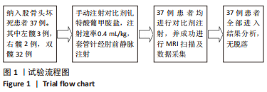
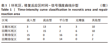
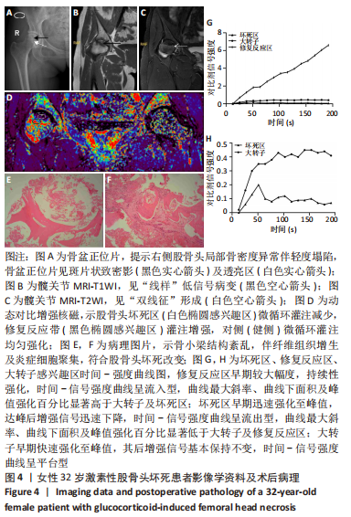
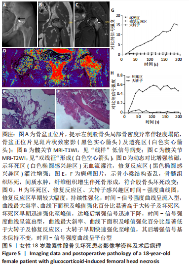
方法:回顾性收集2022-02-16/09-01于江苏省中医院就诊的股骨头坏死37例(69髋)患者的临床资料,其中男29例,女8例;年龄(35.35±12.19)岁(18-65岁)。采用动态对比增强核磁扫描患者双侧股骨头及股骨近端,扫描过程中静脉注射钆特酸葡胺,扫描完成后使用 Geniq软件进行数据后处理得到股骨头微循环灌注参数。在坏死区、修复反应区、大转子处绘制感兴趣区域,获得感兴趣区的时间-信号强度曲线并对其进行分类。使用配对t检验评估坏死区、修复反应区及大转子灌注参数之间的差异以及坏死不同分期股骨头灌注之间的差异;采用χ2检验评估坏死区、修复反应区及大转子时间-强度曲线分型的差异。

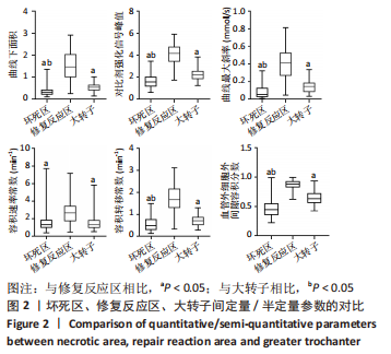
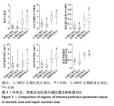
结果与结论:①对比不同区域的灌注参数,坏死区增强信号强度峰值、曲线下面积、最大斜率、容积转移常数、血管外细胞外间隙容积分数小于大转子,差异有显著性意义(P < 0.05),容积速率常数无明显差异(P > 0.05);②修复反应区信号峰值、曲线下面积、最大斜率、容积转移常数、速率转移常数、血管外细胞外间隙比值大于坏死区及大转子,差异有显著性意义(P < 0.05);③ARCOⅡ期坏死区信号峰值及最大斜率大于ARCOⅢ期,差异有显著性意义(P < 0.05);曲线下面积、速率转移常数、容积转移常数、血管外细胞外间隙比值对比无明显差异(P > 0.05);④ARCOⅡ期修复反应区信号峰值、曲线下面积、最大斜率、容积转移常数、速率转移常数、血管外细胞外间隙比值小于ARCOⅢ期,差异有显著性意义(P < 0.05);⑤提示股骨头坏死区微循环灌注速度、血流量及毛细血管渗透性显著降低,修复反应区微循环灌注速度、血流量及血管渗透性较正常区域显著提高,随着塌陷的发生,坏死区血流灌注速度及血流量进一步降低,修复反应区的灌注得到增强。

https://orcid.org/0000-0003-4338-3227 (环大维) 

中国组织工程研究杂志出版内容重点:人工关节;骨植入物;脊柱;骨折;内固定;数字化骨科;组织工程

关键词: 股骨头坏死, 动态对比增强核磁共振, 血流动力学, 灌注, 微循环

Abstract: BACKGROUND: At present, digital subtraction angiography is used in the clinical observation of blood supply of femoral head necrosis, and the patency of the nutrient blood vessels of the femoral head is evaluated by observing the in and out of the contrast agent. Although digital subtraction angiography can visually observe the feeding vessels of the femoral head, it cannot quantify the perfusion. In addition, the collapse of the femoral head is closely related to the prognosis of the disease, but there are few studies on the changes in blood supply in the femoral head during the peri-collapse period.
OBJECTIVE: To compare the differences in microcirculation perfusion in different regions and different stages of femoral head necrosis, summarize the changes in microcirculation perfusion characteristics of femoral head necrosis, and observe the effect of dynamic contrast-enhanced magnetic resonance imaging in evaluating the microcirculation perfusion of the femoral head.
METHODS: From February 16 to September 1, 2022, a total of 37 patients (69 hips) who visited a doctor for femoral head necrosis were retrospectively collected, including 29 males and 8 females; the age was (35.35±12.19) years (range 18-65 years). The patient’s bilateral femoral head and proximal femur were scanned by dynamic contrast-enhanced magnetic resonance imaging, and gadoteric acid meglumine was intravenously injected during the scanning. After scanning, Geniq software was used to post-process the data to obtain femoral head microcirculation perfusion parameters. Regions of interest were drawn at the necrotic zone, repair reaction zone, and greater trochanter, and time-signal intensity curves of the regions of interest were obtained and classified. Paired t-tests were used to evaluate differences between necrotic area, repair reaction area, and greater trochanter perfusion parameters, as well as differences in femoral head perfusion in different stages of necrosis. Differences in necrotic area, repair reaction area and greater trochanter time-intensity curve classification were evaluated using the χ2 test.
RESULTS AND CONCLUSION: (1) Comparing the perfusion parameters in different regions, the peak value of enhanced signal intensity, area under the curve, maximum slope, volume transfer constant, and the median volume fraction of extravascular extracellular space in the necrotic region were lower than those of the greater trochanter, and the differences were statistically significant (P < 0.05). There was no statistically significant difference in the volume rate constant (P > 0.05). (2) The signal peak value, area under the curve, maximum slope, volume transfer constant, rate transfer constant, and the median ratio of extravascular to extracellular space in the repair reaction area were higher than those in the necrotic area and the greater trochanter, and the differences were statistically significant (P < 0.05). (3) The median signal peak value and maximum slope of the necrotic area in ARCO stage II were greater than those in ARCO stage III, and the difference was statistically significant (P < 0.05). There were no significant differences in area under the curve, rate transfer constant, volume transfer constant and the median ratio of extravascular to extracellular space (P > 0.05). (4) The signal peak value, area under the curve, maximum slope, volume transfer constant, rate transfer constant, and ratio of extravascular to extracellular space in the repair reaction zone of the ARCO Stage II were lower than those of the ARCO Stage III, and the difference was statistically significant (P < 0.05). (5) It is concluded that the microcirculation perfusion velocity, blood flow and capillary permeability in the necrotic area of the femoral head were significantly reduced, while the microcirculation perfusion velocity, blood flow and vascular permeability in the repair reaction area were significantly improved compared with those in the normal area. With the occurrence of collapse, the perfusion velocity and blood flow were further reduced, and the perfusion in the repair reaction zone was enhanced.

Key words: femoral head necrosis, dynamic contrast-enhanced magnetic resonance imaging, hemodynamics, perfusion, microcirculation

中图分类号: