中国组织工程研究 ›› 2023, Vol. 27 ›› Issue (6): 860-865.doi: 10.12307/2023.222

• 干细胞外泌体 Stem cell exosomes • 上一篇    下一篇

三种不同来源卵巢颗粒细胞外泌体的提取及鉴定

高  婷1,马小红2,李晓荣2   

  1. 1宁夏医科大学中医学院回医药现代化教育部重点实验室;宁夏医科大学基础医学院生育力保持教育部重点实验室,宁夏回族自治区银川市   750004;2宁夏医科大学总医院生殖医学中心,宁夏回族自治区银川市   750004
  • 收稿日期:2021-12-13 接受日期:2022-02-22 出版日期:2023-02-28 发布日期:2022-08-11
  • 通讯作者: 李晓荣,博士,副主任医师,宁夏医科大学总医院生殖医学中心,宁夏回族自治区银川市 750004
  • 作者简介:高婷,女,1994年生,四川省南充市人,汉族,在读硕士,主要从事女性生殖系统疾病临床与理论研究。
  • 基金资助:
    国家自然科学基金项目(81660806),项目负责人:李晓荣

Extraction and identification of exosomes from three different sources of ovarian granulosa cells

Gao Ting1, Ma Xiaohong2, Li Xiaorong2   

  1. 1School of Traditional Chinese Medicine, Ningxia Medical University, Key Laboratory of Hui Modernization of Medicine, Ministry of Education; Key Laboratory of Fertility Maintenance, Ministry of Education, School of Basic Medicine, Ningxia Medical University, Yinchuan 750004, Ningxia Hui Autonomous Region, China; 2Reproductive Medicine Center, General Hospital of Ningxia Medical University, Yinchuan 750004, Ningxia Hui Autonomous Region, China
  • Received:2021-12-13 Accepted:2022-02-22 Online:2023-02-28 Published:2022-08-11
  • Contact: Li Xiaorong, PhD, Associate chief physician, Reproductive Medicine Center, General Hospital of Ningxia Medical University, Yinchuan 750004, Ningxia Hui Autonomous Region, China
  • About author:Gao Ting, Master candidate, School of Traditional Chinese Medicine, Ningxia Medical University, Key Laboratory of Hui Modernization of Medicine, Ministry of Education; Key Laboratory of Fertility Maintenance, Ministry of Education, School of Basic Medicine, Ningxia Medical University, Yinchuan 750004, Ningxia Hui Autonomous Region, China
  • Supported by:
    the National Natural Science Foundation of China, No.81660806 (to LXR)

摘要:

文题释义:
外泌体:是绝大多数细胞所分泌的具有复杂RNA和蛋白质的小膜泡,其内含物真实地反映了细胞来源和疾病状态;同时它可以作为一种“液体活检”方法,实时评估疾病或人体健康状态;通过其携带和转移的重要信号分子深入了解疾病的发生机制;此外,外泌体还积极参与了细胞间的通讯,并携带多种遗传物质,可通过自分泌或旁分泌被细胞吸收,调节细胞增殖,参与疾病生理病理过程。
颗粒细胞:卵巢颗粒细胞作为卵泡内最大的细胞群,是雌孕激素分泌的主要来源,对卵母细胞以及胚胎发育具有重要的作用,同时它的增殖与分化能力对于卵泡生长发育、排卵、黄体形成和类固醇激素分泌等一系列重要的卵巢功能性活动有着直接的影响。

背景:卵巢颗粒细胞作为卵泡内主要的功能细胞,通过复杂的缝隙连接与卵母细胞进行细胞间的信息物质交流,调控着卵母细胞的生长与成熟;随着外泌体研究的深入,发现其积极参与了细胞间的通讯,并携带多种遗传物质,可通过自分泌或旁分泌方式被细胞吸收,调节细胞增殖,参与疾病生理病理过程。由此可见,卵巢颗粒细胞的外泌体分泌能力及提取鉴定对于研究女性生殖具有重要的意义。
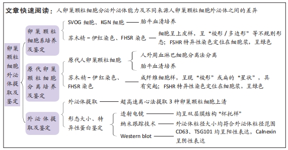
目的:探讨人卵巢颗粒细胞分泌外泌体的能力及不同来源人卵巢颗粒细胞外泌体之间的差异。
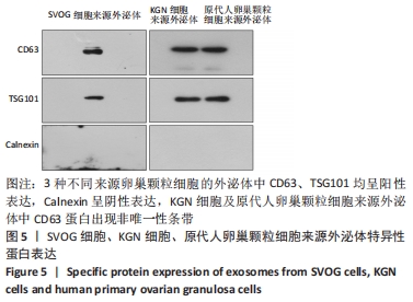
方法:体外培养SVOG细胞(人卵巢颗粒细胞系)、KGN细胞(人卵巢颗粒细胞瘤细胞)、人原代卵巢颗粒细胞;采用超高速离心法分别从3种不同来源卵巢颗粒细胞上清中分离提取外泌体,通过透射电子显微镜、纳米跟踪技术、蛋白质印迹法分别鉴定其形态结构、粒径大小、阳性相关特异性蛋白CD63、TSG101及阴性标记蛋白Calnexin的表达。
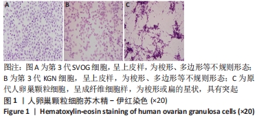
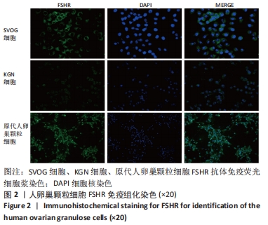
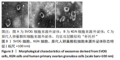
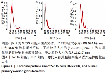
结果与结论:①SVOG细胞、KGN细胞、人原代卵巢颗粒细胞的细胞核均为圆形或椭圆形,核大,核仁部分蓝染,细胞浆呈淡红色;SVOG细胞、KGN细胞呈长梭形或多边形,形态不规则,人原代卵巢颗粒细胞多呈突起或梭形,细胞伪足细长,相互紧密交织;②3种不同来源人卵巢颗粒细胞免疫荧光均阳性表达FSHR,且阳性表达率> 95%;③3种不同细胞上清液分离的外泌体均呈双层膜结构杯托样;④纳米跟踪分析SVOG细胞、KGN细胞、人原代卵巢颗粒细胞来源外泌体的平均粒径分别为(183.5±4.9),(125.3±1.0),(171.1±2.0) nm,其中人原代卵巢颗粒细胞来源外泌体的分泌水平最高,KGN细胞来源外泌体的粒径最小,SVOG细胞来源外泌体大小较接近人原代卵巢颗粒细胞来源外泌体;⑤蛋白质印迹法检测显示3种不同来源卵巢颗粒细胞来源外泌体特异性相关蛋白CD63、TSG101均呈阳性表达,而Calnexin不表达;⑥结果表明,采用超高速离心法能够成功分离提取不同卵巢颗粒细胞来源外泌体,KGN细胞来源外泌体粒径最小,SVOG细胞及人原代卵巢颗粒细胞来源外泌体粒径较接近,以人原代卵巢颗粒细胞来源外泌体的分泌水平最高。

https://orcid.org/0000-0002-5746-1727 (高婷)

中国组织工程研究杂志出版内容重点:干细胞;骨髓干细胞;造血干细胞;脂肪干细胞;肿瘤干细胞;胚胎干细胞;脐带脐血干细胞;干细胞诱导;干细胞分化;组织工程

关键词: 颗粒细胞, 外泌体, 透射电镜, 纳米粒子示踪分析, 蛋白质印迹法

Abstract: BACKGROUND: Ovarian granulosa cells, as the main functional cells in ovarian follicles, communicate with oocytes through complex gap junctions, and regulate the growth and maturation of oocytes. With the deepening of exosome research, it is found that it actively participates in the communication between cells, and carries a variety of genetic materials, which can be absorbed by cells through autocrine or paracrine, regulate cell proliferation, and participate in the physiological and pathological processes of diseases. It can be seen that the exosome secretion capacity and extraction and identification of ovarian granulosa cells are of great significance for the study of female reproduction.  
OBJECTIVE: To explore the ability of human ovarian granulosa cells to isolate exosomes and the differences between exosomes from human ovarian granulosa cells of different origins.
METHODS:  SVOG (human ovarian granulosa cell line), KGN (human ovarian granulosa cell tumor cells) and human primary ovarian granulosa cells were cultured in vitro. Exosomes were isolated from the supernatants of the three different cells by ultra-high speed centrifugation. The morphological structures, particle sizes, and the expression of CD63, tumor susceptibility gene (TSG101) and calnexin were measured by transmission electron microscopy, nanoparticle tracer technique, and western blot assay, respectively.  
RESULTS AND CONCLUSION: (1) The nuclei of SVOG cells, KGN cells and human primary ovarian granulosa cells were round or oval with large nuclei, blue-stained nucleosomes and light red cytoplasm. SVOG cells and KGN cells were long spindle-shaped or polygonal with irregular morphology, and human primary ovarian granulosa cells were mostly protruding or spindle-shaped with elongated pseudopodia and closely intertwined with each other. (2) Immunofluorescence from three different sources of human ovarian granulosa cells was positive for FSHR expression, and the positive expression rate was greater than 95%. (3) The morphology of the exosomes isolated from the supernatants of the three different cells presented a cup-like bilayer structure. (4) Nanoparticle tracer technique showed that the mean particle sizes of exosomes from SVOG cells, KGN cells and human primary ovarian granulosa cells were (183.5±4.9) nm, (125.3±1.0) nm, and (171.1±2.0) nm, respectively. Among them, human primary ovarian granulosa cell-derived exosomes had the highest secretion levels. The exosomes from KGN cells had the smallest particle size, and the exosomes from SVOG cells were relatively similar in size to those from human primary ovarian granulosa cells. (5) Western blot assasy results showed that the exosomes from three different sources of ovarian granulosa cells were positive for the specific protein CD63 and the tumor susceptibility gene (TSG101) but negative for Calnexin. (6) These findings confirm that the ultra-high speed centrifugation method can successfully isolate and extract ovarian granulosa cell exosomes, among which KGN cell-derived exosomes have the smallest particle size; the particle sizes of exosomes derived from SVOG cells and human primary ovarian granulosa cells are similar; human primary ovarian granulosa cells have the strongest ability to secrete exosomes.

Key words: granulosa cell, exosome, transmission electron microscopy, nanoparticle tracer analysis, western blot assay

中图分类号: