[1] VANTYGHEM MC, DE KONING EJP, PATTOU F, et al. Advances in β-cell replacement therapy for the treatment of type 1 diabetes. Lancet. 2019;394(10205):1274-1285.
[2] SCHASCHKOW A, SIGRIST S, MURA C, et al. Glycaemic control in diabetic rats treated with islet transplantation using plasma combined with hydroxypropylmethyl cellulose hydrogel. Acta Biomater. 2020; 102(259-272.
[3] SHAPIRO AM, POKRYWCZYNSKA M, RICORDI C. Clinical pancreatic islet transplantation. Nat Rev Endocrinol. 2017;13(5):268-277.
[4] LLACUA LA, FAAS MM, DE VOS P. Extracellular matrix molecules and their potential contribution to the function of transplanted pancreatic islets. Diabetologia. 2018;61(6):1261-1272.
[5] SMINK AM, DE VOS P. Therapeutic Strategies for Modulating the Extracellular Matrix to Improve Pancreatic Islet Function and Survival After Transplantation. Curr Diab Rep. 2018;18(7):39.
[6] ZHANG F, ZHENG L, CHENG S, et al. Comparison of the Interactions of Different Growth Factors and Glycosaminoglycans. Molecules. 2019; 24(18):3360.
[7] LI D, ZHANG W, CHEN X, et al. Proteomic profiling of MIN6 cell-derived exosomes. J Proteomics. 2020;224:103841.
[8] YIN H, YAN H, QIN C, et al. Protective effect of fermented Diospyros lotus L. extracts against the high glucose-induced apoptosis of MIN6 cells. J Food Biochem. 2021;45(4):e13685.
[9] HU S, KUWABARA R, DE HAAN B, et al. Acetate and Butyrate Improve β-cell Metabolism and Mitochondrial Respiration under Oxidative Stress. Int J Mol Sci. 2020;21(4):1542.
[10] BEENKEN-ROTHKOPF L, KARFELD-SULZER L, DAVIS N, et al. The incorporation of extracellular matrix proteins in protein polymer hydrogels to improve encapsulated beta-cell function. Ann Clin Lab Sci. 2013;43(2):111-121.
[11] LIM D, ANTIPENKO S, VINES J, et al. Improved MIN6 β-cell function on self-assembled peptide amphiphile nanomatrix inscribed with extracellular matrix-derived cell adhesive ligands. Macromol Biosci. 2013;13(10):1404-1412.
[12] 潘钰.富血小板血浆治疗安全,有效[J].江苏卫生保健,2019(4):1.
[13] 吕敏,裴国献,刘勇,等.富血小板血浆的制备现状及研究进展[J].现代生物医学进展,2013,13(27):5.
[14] ANITUA E, PRADO R, ORIVE G. Allogeneic Platelet-Rich Plasma: At the Dawn of an Off-the-Shelf Therapy? Trends Biotechnol. 2017;35(2):91-93.
[15] BARIA M, VASILEFF W, MILLER M, et al. Cellular Components and Growth Factor Content of Platelet-Rich Plasma With a Customizable Commercial System. Am J Sports Med. 2019;47(5):1216-1222.
[16] OUDELAAR B, PEERBOOMS J, HUIS IN ‘T VELD R , et al. Concentrations of Blood Components in Commercial Platelet-Rich Plasma Separation Systems: A Review of the Literature. Am J Sports Med. 2019;47(2): 479-487.
[17] EPPLEY B, WOODELL J, HIGGINS JJP, et al. Platelet quantification and growth factor analysis from platelet-rich plasma: implications for wound healing. Plast Reconstr Surg. 2004;114(6):1502-1508.
[18] PULCINI S, MEROLLE L, MARRACCINI C, et al. Apheresis Platelet Rich-Plasma for Regenerative Medicine: An In Vitro Study on Osteogenic Potential. Int J Mol Sci. 2021;22(16):8764.
[19] WEI S, XU P, YAO Z, et al. A composite hydrogel with co-delivery of antimicrobial peptides and platelet-rich plasma to enhance healing of infected wounds in diabetes. Acta Biomater. 2021;124:205-218.
[20] SAMBERG M, STONE R, NATESAN S, et al. Platelet rich plasma hydrogels promote in vitro and in vivo angiogenic potential of adipose-derived stem cells. Acta Biomater. 2019;87:76-87.
[21] DE ANGELIS B, D’AUTILIO M, ORLANDI F, et al. Wound Healing: In Vitro and In Vivo Evaluation of a Bio-Functionalized Scaffold Based on Hyaluronic Acid and Platelet-Rich Plasma in Chronic Ulcers. J Clin Med. 2019;8(9):1486.
[22] BELLIS SJB. Advantages of RGD peptides for directing cell association with biomaterials. Biomaterials. 2011;32(18):4205-4210.
[23] ROSE J, PACELLI S, HAJ A, et al. Gelatin-Based Materials in Ocular Tissue Engineering. Materials(Basel). 2014;7(4):3106-3135.
[24] YUE K, TRUJILLO-DE SANTIAGO G, ALVAREZ MM, et al. Synthesis, properties, and biomedical applications of gelatin methacryloyl (GelMA) hydrogels. Biomaterials. 2015;73:254-271.
[25] SHIN SR, ZIHLMANN C, AKBARI M, et al. Reduced Graphene Oxide-GelMA Hybrid Hydrogels as Scaffolds for Cardiac Tissue Engineering. Small. 2016;12(27):3677-3689.
[26] MODARESIFAR K, HADJIZADEH A, NIKNEJAD HJAC, et al. Design and fabrication of GelMA/chitosan nanoparticles composite hydrogel for angiogenic growth factor delivery. Artif Cells Nanomed Biotechnol. 2018;46(8):1799-1808.
[27] WU S, WANG L, FANG Y, et al. Advances in Encapsulation and Delivery Strategies for Islet Transplantation. Adv Healthc Mater. 2021;10(20): e2100965.
[28] TERAMURA Y, OOMMEN O, OLERUD J, et al. Microencapsulation of cells, including islets, within stable ultra-thin membranes of maleimide-conjugated PEG-lipid with multifunctional crosslinkers. Biomaterials. 2013;34(11):2683-2693.
[29] URBANCZYK M, ZBINDEN A, LAYLAND S, et al. Controlled Heterotypic Pseudo-Islet Assembly of Human β-Cells and Human Umbilical Vein Endothelial Cells Using Magnetic Levitation. Tissue Eng Part. 2020;26: 387-399.
[30] ZHU Q, LU C, JIANG X, et al. Using Recombinant Human Collagen With Basic Fibroblast Growth Factor to Provide a Simulated Extracellular Matrix Microenvironment for the Revascularization and Attachment of Islets to the Transplantation Region. Front Pharmacol. 2019;10:1536.
[31] ZHONG Z, BALYAN A, TIAN J, et al. Bioprinting of dual ECM scaffolds encapsulating limbal stem/progenitor cells in active and quiescent statuses. Biofabrication. 2021;13(4):044101.
[32] JIN R, CUI Y, CHEN H, et al. Three-dimensional bioprinting of a full-thickness functional skin model using acellular dermal matrix and gelatin methacrylamide bioink. Acta Biomater. 2021;131:248-261.
[33] VAN DEN BULCKE A, BOGDANOV B, DE ROOZE N, et al. Structural and rheological properties of methacrylamide modified gelatin hydrogels. Biomacromolecules. 2000;1(1):31-38.
[34] SUN X, MA Z, ZHAO X, et al. Three-dimensional bioprinting of multicell-laden scaffolds containing bone morphogenic protein-4 for promoting M2 macrophage polarization and accelerating bone defect repair in diabetes mellitus. Bioact Mater. 2020;6(3):757-769.
[35] EVERTS P, ONISHI K, JAYARAM P, et al. Platelet-Rich Plasma: New Performance Understandings and Therapeutic Considerations in 2020. Int J Mol Sci. 2020;21(20):7794.
[36] FADADU PP, MAZZOLA AJ, HUNTER CW, et al. Review of concentration yields in commercially available platelet-rich plasma (PRP) systems: a call for PRP standardization. Reg Anesth Pain Med. 2019 :rapm-2018-100356.
[37] KLEIN M, KäMMERER P, SCHOLZ T, et al. Modulation of platelet activation and initial cytokine release by alloplastic bone substitute materials. Clin Oral Implants Res. 2010;21(3):336-345.
[38] HUBER S, CUNHA JúNIOR J, MONTALVãO S, et al. In vitro study of the role of thrombin in platelet rich plasma (PRP) preparation: utility for gel formation and impact in growth factors release. J Stem Cells Regen Med. 2016;12(1):2-9.
[39] GAO X, GAO L, GROTH T, et al. Fabrication and properties of an injectable sodium alginate/PRP composite hydrogel as a potential cell carrier for cartilage repair. J Biomed Mater Res A. 2019;107(9):2076-2087.
[40] ENDERAMI SE, MORTAZAVI Y, SOLEIMANI M, et al. Generation of Insulin-Producing Cells From Human-Induced Pluripotent Stem Cells Using a Stepwise Differentiation Protocol Optimized With Platelet-Rich Plasma. J Cell Physiol. 2017;232(10):2878-2886.
[41] ANITUA E, DE LA SEN-CORCUERA B, ORIVE G, et al. Progress in the use of plasma rich in growth factors in ophthalmology: from ocular surface to ocular fundus. Expert Opin Biol Ther. 2022;22(1):31-45.
[42] BYNIGERI RR, MITNALA S, TALUKDAR R, et al. Pancreatic stellate cell-potentiated insulin secretion from Min6 cells is independent of interleukin 6-mediated pathway. J Cell Biochem. 2020;121(1):840-855.
[43] FURUKAWA A, TANAKA A, YAMAGUCHI S, et al. Using recombinase-mediated cassette exchange to engineer MIN6 insulin-secreting cells based on a newly identified safe harbor locus. J Diabetes Investig. 2021;12(12):2129-2140.
[44] SCHULTZ J, WARKUS J, WOLKE C, et al. MiD51 Is Important for Maintaining Mitochondrial Health in Pancreatic Islet and MIN6 Cells. Front Endocrinol (Lausanne). 2020;11:232.
[45] BLESIA V, PATEL VB, AL-OBAIDI H, et al. Excessive Iron Induces Oxidative Stress Promoting Cellular Perturbations and Insulin Secretory Dysfunction in MIN6 Beta Cells. Cells. 2021;10(5):1141.
[46] 石红梅,张英兰,侯小红,等.外观异常的血清标本对生化结果的影响[J].实用医技杂志,2003,10(12):1495-1495.
[47] SIBORO S, ANUGRAH D, RAMESH K, et al. Tunable porosity of covalently crosslinked alginate-based hydrogels and its significance in drug release behavior. Carbohydr Polym. 2021;260:117779.
[48] 姜铧,张洹,李东升,等.PDX1与糖尿病[J].湖北医药学院学报, 2008,27(1):81-84.
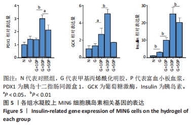
[49] 邹大进.血糖稳态调控的核心酶:葡萄糖激酶及其家族[J].中华糖尿病杂志,2019,11(10):3.
|