中国组织工程研究 ›› 2023, Vol. 27 ›› Issue (8): 1179-1186.doi: 10.12307/2022.1001

• 组织构建实验造模 experimental modeling in tissue construction • 上一篇    下一篇

Zeste基因抑制子基因12在糖尿病肾脏病模型大鼠肾组织中的表达

赵 璐1,赵逸菲2,高 达1,刘艳芳3,付婷婷1,徐江雁4   

  1. 河南中医药大学第三附属医院,1内分泌科,3肾病科,河南省郑州市  450008;2河南中医药大学龙子湖校区,河南省郑州市  450046;4河南中医药大学,河南省郑州市  450046
  • 收稿日期:2021-10-25 接受日期:2022-01-13 出版日期:2023-03-18 发布日期:2022-07-27
  • 通讯作者: 徐江雁,博士,教授,副校长,河南中医药大学,河南省郑州市 450046
  • 作者简介:赵璐,女,1974年生,2011年山东中医药大学毕业,博士,副主任中医师,主要从事内分泌代谢研究。
  • 基金资助:
    国家重点研发计划项目(2020YFE0201800),项目负责人:徐江雁

Expression of suppressor of Zeste 12 in kidney tissues of rats with diabetic nephropathy

Zhao Lu1, Zhao Yifei2, Gao Da1, Liu Yanfang3, Fu Tingting1, Xu Jiangyan4   

  1. 1Department of Endocrinology, 3Department of Nephrology, the Third Affiliated Hospital of Henan University of Chinese Medicine, Zhengzhou 450008, Henan Province, China; 2Longzi Hu Campus, Henan University of Chinese Medicine, Zhengzhou 450046, Henan Province, China; 4Henan University of Chinese Medicine, Zhengzhou 450046, Henan Province, China
  • Received:2021-10-25 Accepted:2022-01-13 Online:2023-03-18 Published:2022-07-27
  • Contact: Xu Jiangyan, MD, Professor, Henan University of Chinese Medicine, Zhengzhou 450046, Henan Province, China
  • About author:Zhao Lu, MD, Associate chief physician, Department of Endocrinology, the Third Affiliated Hospital of Henan University of Chinese Medicine, Zhengzhou 450008, Henan Province, China
  • Supported by:
    the National Key R&D Program Project of China, No. 2020YFE0201800 (to XJY)

摘要:

文题释义:
糖尿病肾脏病:是由糖尿病引起的肾脏疾病,属于糖尿病最严重的微血管并发症之一,已成为世界终末期肾脏病的第2位原因,仅次于肾小球肾炎。肾小管间质纤维化是导致糖尿病终末期肾功能衰竭的主要原因之一。
SUZ12:是Polycomb 抑制复合物 2(PRC2)的核心蛋白,是一种组蛋白甲基化转移酶,指导组蛋白3赖氨酸 27 (H3K27Me3)的甲基化和基因沉默。研究表明,SUZ12 蛋白在包括肾癌在内的多种肿瘤中过度表达,促进肿瘤细胞迁移、侵袭以及上皮间质转化。其中,上皮细胞-间充质细胞转化是导致糖尿病肾脏病肾脏纤维化发生的机制之一,因此SUZ12可能在糖尿病肾脏病的发展过程中起着至关重要的作用。

背景:Zeste基因抑制子基因12(Suppressor of zeste gene 12,Suz12)可参与肾小管上皮细胞的上皮间质转化。
目的:探讨Suz12对糖尿病肾脏病进程的影响及其相关作用机制。
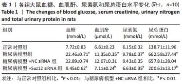
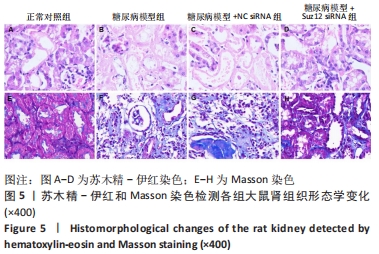
方法:①细胞实验:将大鼠肾小管上皮细胞分为正常对照组(葡萄糖5.5 mmol/L)、高糖组(葡萄糖30 mmol/L)和高渗组(葡萄糖5.5 mmol/L+甘露醇24.5 mmol/L);将100 nmol/L Suz12小干扰RNA(siRNA)及其阴性对照(NC siRNA)转染至高糖培养的肾小管上皮细胞中后48 h,采用Western blotting、CCK-8和流式细胞仪检测Suz12、Ⅳ型胶原蛋白、α-平滑肌肌动蛋白和E-cadherin的蛋白表达、细胞增殖和凋亡。采用染色质免疫共沉淀检测Suz12和金属蛋白酶组织抑制因子3的结合;金属蛋白酶组织抑制因子3表达抑制与组蛋白H3K27me水平增加有关,以甲基化特异性PCR法检测金属蛋白酶组织抑制因子3组蛋白H3K27me3甲基化水平;采用Wnt/β-catenin通路激活剂TDZD-8(10 μmol/L,1 h)处理转染Suz12 siRNA的肾小管上皮细胞,以验证Wnt通路的激活是否影响干扰Suz12对细胞损伤的作用。②动物实验:采用腹腔注射60 mg/kg链脲佐菌素构建糖尿病大鼠模型,并通过尾静脉注射0.1 mL含3×10⁸ PFU空腺病毒载体(NC siRNA)或Suz12 siRNA腺病毒(Suz12 siRNA)的PBS;实验结束后收集血清、尿液和肾脏组织,采用全自动生化分析仪检测血糖、血肌酐、尿氮素、尿总蛋白水平;苏木精-伊红和Masson 染色观察肾脏组织形态学变化;Western blotting检测肾脏组织中相关蛋白的表达水平。
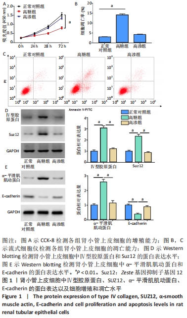
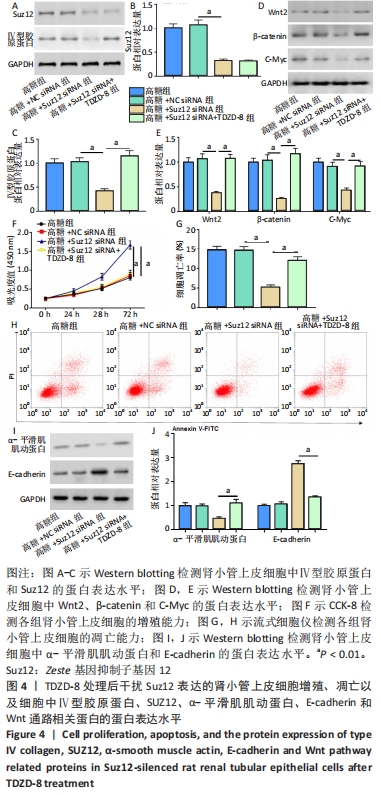
结果与结论:①与正常对照组相比,高糖组肾小管上皮细胞中Suz12、Ⅳ型胶原蛋白和α-平滑肌肌动蛋白蛋白表达水平明显升高,E-cadherin蛋白表达水平明显降低,细胞增殖减少,凋亡率增加;而转染Suz12 siRNA可逆转高糖处理对肾小管上皮细胞的损伤;②与高糖+Suz12 siRNA组相比,TDZD-8处理可逆转Suz12敲低对肾小管上皮细胞中Ⅳ型胶原蛋白、α-平滑肌肌动蛋白、E-cadherin和Wnt通路相关蛋白表达水平、细胞增殖和凋亡的影响;③Suz12可与金属蛋白酶组织抑制因子3结合,显示金属蛋白酶组织抑制因子3存在H3K27me3修饰,且敲低Suz12可降低高糖诱导的肾小管上皮细胞中金属蛋白酶组织抑制因子3甲基化水平;④与糖尿病组相比,Suz12 siRNA组大鼠血糖、血肌酐、尿氮素、尿总蛋白水平明显降低,肾脏组织病理变化和纤维增生得到缓解,Ⅳ型胶原蛋白、Suz12和Wnt通路相关蛋白表达水平显著降低,E-cadherin表达水平明显升高;⑤结果表明,Suz12在糖尿病大鼠中可能通过促进金属蛋白酶组织抑制因子3甲基化参与糖尿病肾脏病发展。
缩略语:Zeste基因抑制子基因12:Suppressor of zeste gene 12,Suz12;金属蛋白酶组织抑制因子3:tissue inhibitor of metalloproteinases-3,TIMP3

https://orcid.org/0000-0002-4505-6361 (赵璐)

中国组织工程研究杂志出版内容重点:组织构建;骨细胞;软骨细胞;细胞培养;成纤维细胞;血管内皮细胞;骨质疏松;组织工程

关键词: 糖尿病肾脏病, Suz12, TIMP3, 甲基化, Wnt/β-catenin信号通路

Abstract: BACKGROUND: Suppressor of zeste 12 (Suz12) can participate in the epithelial-mesenchymal transition of tubular epithelial cells.
OBJECTIVE: To investigate the effect of Suz12 on the progression of diabetic nephropathy and its related mechanism. 
METHODS: (1) Cell experiment: Rat renal tubular epithelial cells were set as normal control group (glucose 5.5 mmol/L), high glucose group (glucose 30 mmol/L), and hypertonic group (glucose 5.5 mmol/L+mannitol 24.5 mmol/L). After transfection of 100 nmol/L Suz12 small interfering RNA (siRNA) and its negative control (NC siRNA) into renal tubular epithelial cells cultured in high glucose, western blot, cell counting kit-8, and flow cytometry were used to detect the protein expression of type IV collagen, Suz12, α-smooth muscle actin, and E-cadherin, cell proliferation and apoptosis, respectively. Subsequently, chromatin immunoprecipitation was used to detect the binding of Suz12 and tissue inhibitor of metalloproteinases-3 (TIMP3). The inhibition of TIMP3 expression is associated with the increase of trimethylation of lysine 27 on histone 3 (H3K27me). Methylation-specific PCR method was used to detect the methylation level of TIMP3 histone H3K27me3. Renal tubular epithelial cells were treated with Wnt/β-catenin pathway activator TDZD-8 (10 μmol/L; 1 hour) to verify whether the activation of Wnt pathway influences the effects of Suz12 on cell injury. (2) Animal experiment: A diabetic rat model was established by intraperitoneal injection of 60 mg/kg streptozotocin, and then 0.1 mL of PBS solution containing 3×10⁸ PFU empty adenovirus vector (NC siRNA) or Suz12 siRNA adenovirus (Suz12 siRNA) was injected through the tail vein. After the experiment, serum, urine, and kidney tissues were collected, and the contents of blood glucose, serum creatinine, urinary nitrogen and total urinary protein were detected by an automatic biochemical analyzer. Hematoxylin-eosin and Masson staining were used to observe the renal histomorphological changes. Western blot assay was used to detect the expressions of related proteins in kidney tissues.  
RESULTS AND CONCLUSION: Compared with the normal control group, the expression levels of Suz12, type IV collagen, α-smooth muscle actin in the renal tubular epithelial cells were significantly increased, E-cadherin expression was decreased, cell proliferation was decreased, and the apoptotic rate was increased in the high glucose group, whilst transfection of Suz12 siRNA could reverse the damage of high glucose treatment to the renal tubular epithelial cells. Compared with the high glucose+Suz12 siRNA group, TDZD-8 treatment could reverse the effects of Suz12 knockdown on type IV collagen, α-smooth muscle actin, E-cadherin and Wnt pathway related protein expression levels, cell proliferation and apoptosis of renal tubular epithelial cells. Suz12 could specifically bind to TIMP3 protein, indicating that TIMP3 is modified by H3K27me3, and knockdown of Suz12 reduced the level of TIMP3 methylation in high glucose-stimulated renal tubular epithelial cells. Compared with the diabetic model group, serum glucose, serum creatinine, urinary nitrogen, and urinary total protein contents were significantly decreased, renal histopathological changes and fibrous hyperplasia were alleviated, and Suz12, collagen 4, α-smooth muscle actin, E-cadherin and Wnt pathway related protein expression levels were significantly decreased in the diabetic model+Suz12 siRNA group. The above results indicate that Suz12 may be involved in the development of diabetic nephropathy by promoting the methylation of TIMP3 in diabetic rats.

Key words: diabetic nephropathy, Suz12, TIMP3, methylation, Wnt/β-catenin signaling pathway

中图分类号: