[1] Orthopedic Implants Market Analysis,By Application (Spinal fusion Long bone Foot & Ankle Craniomaxillofacial Joint replacement Dental) And Segment Forecasts To 2024. GRAND VIEW RESEARCH,2016.
[2] FESTAS A, RAMOS A, DAVIM J. Medical devices biomaterials–a review.P I Mech ENGL-J MAT.2020;234(1):218-228.
[3] SONG Y, XU D, YANG R, et al. Theoretical study of the effects of alloying elements on the strength and modulus of β-type bio-titanium alloys.Mater Sci Eng A.1999;260(1-2):269-274.
[4] GEPREEL M AH, NIINOMI M. Biocompatibility of Ti-alloys for long-term implantation. J Mech Behav Biomed Mater.2013;20:407-415.
[5] LÜTJERING G, WILLIAMS JC. Titanium. Springer Science & Business Media,2007.
[6] ZPRIANO S, YAMAGUCHI S, BAINO F, et al. A critical review of multifunctional titanium surfaces:New frontiers for improving osseointegration and host response,avoiding bacteria contamination.Acta Biomater.2018;79:1-22.
[7] ZHAO B, VAN DER MEI HC, SUBBIAHDOSS G, et al. Soft tissue integration versus early biofilm formation on different dental implant materials.Dent mater.2014;30(7):716-727.
[8] LI Y, YANG Y, LI R, et al. Enhanced antibacterial properties of orthopedic implants by titanium nanotube surface modification:a review of current techniques.Int J Nanomed.2019;14:7217.
[9] ABAD CL, HALEEM A. Prosthetic Joint Infections:an Update.Curr Infect Dis Rep.2018;20(7):15.
[10] WANG Q, HUANG JY, LI HQ, et al. TiO2 nanotube platforms for smart drug delivery:a review.Int J Nanomed.2016;11:4819.
[11] BARIANA M, KAIDONIS JA, LOSIC D, et al. Titania nanotube-based protein delivery system to inhibit cranial bone regeneration in Crouzon model of craniosynostosis.Int J Nanomed.2019;14:6313.
[12] SZCZEŚ A, HOŁYSZ L, CHIBOWSKI E. Synthesis of hydroxyapatite for biomedical applications.Adv Colloid Interface Sci.2017;249:321-330.
[13] XIONG L, ZENG J, YAO A, et al. BMP2-loaded hollow hydroxyapatite microspheres exhibit enhanced osteoinduction and osteogenicity in large bone defects.Int J Nanomed.2015;10:517.
[14] YANG H, GAO H, WANG Y. Hollow hydroxyapatite microsphere:a promising carrier for bone tissue engineering.J Microencapsulation. 2016;33(5):421-426.
[15] MUNIR MU, IHSAN A, SARWAR Y, et al. Hollow mesoporous hydroxyapatite nanostructures:smart nanocarriers with high drug loading and controlled releasing features.Int J Pharmaceut.2018; 544(1):112-120.
[16] LAI W, CHEN C, REN X, et al. Hydrothermal fabrication of porous hollow hydroxyapatite microspheres for a drug delivery system.Mater Sci Eng C.2016;62:166-172.
[17] MONDAL S, DOROZHKIN SV, PAL U. Recent progress on fabrication and drug delivery applications of nanostructured hydroxyapatite.Wiley Interdiscip Rev Nanomed Nanobiotechnol.2018;10(4):e1504.
[18] WANG K, WANG Y, ZHAO X, et al. Sustained release of simvastatin from hollow carbonated hydroxyapatite microspheres prepared by aspartic acid and sodium dodecyl sulfate.Mater Sci Eng C.2017;75:565-571.
[19] WEERASURIYA D, WIJESINGHE W, RAJAPAKSE R. Encapsulation of anticancer drug copper bis in hydroxyapatite for pH-sensitive targeted delivery and slow release.Mater Sci Eng C.2017;71:206-213.
[20] LIU J, QIAO SZ, LIU H, et al. Extension of the Stöber method to the preparation of monodisperse resorcinol-formaldehyde resin polymer and carbon spheres.Angew Chem Int Ed.2011;50(26):5947-5951.
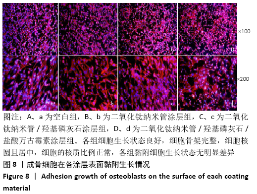
[21] 雷则鸣,张杭州,田昂,等.纳米羟基磷灰石/大管径TiO2纳米管复合涂层促成骨[J].中国组织工程研究,2017,21(14):2186-2191.
[22] 张杭州,田昂,梁庆威,等.万古霉素/羟基磷灰石/二氧化钛纳米管的抗菌性能[J].中国组织工程研究,2016,20(25):3732-3737.
[23] JING W, HUANG SP, KUN H, et al. Effect of cetyltrimethylammonium bromide on morphology and porous structure of mesoporous hydroxyapatite.Trans Nonferrous Met Soc China.2015;25(2):483-489.
[24] 10993-12 ISO. Biological evaluation of medical devices:Sample preparation and reference materials.
[25] 周建军,乐秀芳.评价抗癌物质活性的改良MTT方法[J].中国医药工业杂志,1993,24(10):455-457.
[26] 10993-5 ISO. Biological evaluation of medical devices:Tests for in vitro cytotoxicity,2009.
[27] ALT V. Antimicrobial coated implants in trauma and orthopaedics-A clinical review and risk-benefit analysis.Injury.2017;48(3):599-607.
[28] BJURSTEN LM, RASMUSSON L, OH S, et al. Titanium dioxide nanotubes enhance bone bonding in vivo.Biomed Mater Res P A-J.2010;92(3): 1218-1224.
[29] AL-MOBARAK N, AL-SWAYIH A. Development of titanium surgery implants for improving osseointegration through formation of a titanium nanotube layer.Int J Electrochem Sci.2014;9:32-45.
[30] IVANOVA EP, TRUONG VK, WANG JY, et al. Impact of nanoscale roughness of titanium thin film surfaces on bacterial retention.Langmuir. 2010;26(3):1973-1982.
[31] DENG Y, SUN M, SHAEVITZ J W. Measuring peptidoglycan elasticity and stress-stiffening of live bacterial cells.Biophys J.2011;100(3): 514a-515a.
[32] OLIVEIRA W F, ARRUDA I R, SILVA GM, et al. Functionalization of titanium dioxide nanotubes with biomolecules for biomedical applications.Mater Sci Eng:C.2017;81:597-606.
[33] BARFEIE A, WILSON J, REES J. Implant surface characteristics and their effect on osseointegration.Br dent J.2015;218(5):E9.
[34] ABDELTAWAB A, SHOEIB M, MOHAMED S. Electrophoretic deposition of hydroxyapatite coatings on titanium from dimethylformamide suspensions.Surf Coat Technol.2011;206(1):43-50.
[35] PAWLIK A, REHMAN M AU, NAWAZ Q, et al. Fabrication and characterization of electrophoretically deposited chitosan-hydroxyapatite composite coatings on anodic titanium dioxide layers.Electrochim Acta.2019;307:465-473.
[36] PANTOVIĆ PAVLOVIĆ MR, PAVLOVIĆ MM, ERAKOVIĆ SG, et al. Novel in-situ synthesis of hydroxyapatite/titanium oxide composite coatings on titanium by simultaneous anodization/anaphoretic electrodeposition.Eng Ecology Mater P Indust.2019;3:11-13.
[37] AHMADI S, MOHAMMADI I, SADRNEZHAAD S. Hydroxyapatite based and anodic Titania nanotube biocomposite coatings: Fabrication, characterization and electrochemical behavior.Surf Coat Tech. 2016; 287:67-75.
[38] IONITA D, BAJENARU-GEORGESCU D, TOTEA G, et al. Activity of vancomycin release from bioinspired coatings of hydroxyapatite or TiO2 nanotubes.Int J Pharm.2017;517(1-2):296-302.
[39] GOUDARZI M, BATMANGHELICH F, AFSHAR A, et al. Development of electrophoretically deposited hydroxyapatite coatings on anodized nanotubular TiO2 structures:corrosion and sintering temperature.Appl Surf Sci.2014;301:250-257.
[40] MATHEW D, BHARDWAJ G, WANG Q, et al. Decreased Staphylococcus aureus and increased osteoblast density on nanostructured electrophoretic-deposited hydroxyapatite on titanium without the use of pharmaceuticals.Int J Nanomed.2014;9:1775.
[41] SADRNEZHAAD S, ABBASPOUR S. Loading drug on nanostructured Ti6Al4V-HA for implant applications.Int J Eng.2018;31(8):1159-1165.
[42] ZHANG X, ZHANG Y, YATES MZ. Hydroxyapatite Nanocrystal Deposited Titanium Dioxide Nanotubes Loaded with Antibiotics for Combining Biocompatibility and Antibacterial Properties.MRS Adv. 2018; 3(30): 1703-1709.
[43] LIU J X, BRAVO D, BUZA J, et al. Topical vancomycin and its effect on survival and migration of osteoblasts,fibroblasts,and myoblasts:An in vitro study.J Orthop Sci.2018;15(1):53-58.
[44] STEWART C, AKHAVAN B, WISE SG, et al. A review of biomimetic surface functionalization for bone-integrating orthopedic implants: Mechanisms,current approaches,and future directions.Prog Mater Sci. 2019;100588.
|